. 2000 Jan;119(1):140-7.
doi: 10.1046/j.1365-2249.2000.01098.x.

Spontaneous and oxidative stress-induced programmed cell death in lymphocytes from patients with ataxia telangiectasia (AT)

Affiliations

Spontaneous and oxidative stress-induced programmed cell death in lymphocytes from patients with ataxia telangiectasia (AT)

R Schubert et al. Clin Exp Immunol. 2000 Jan.

Abstract

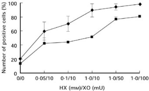
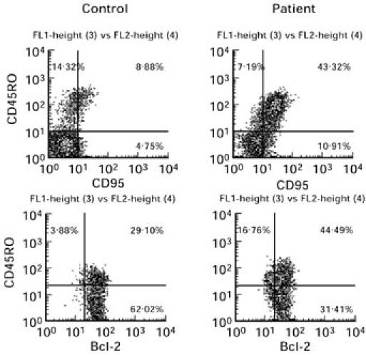
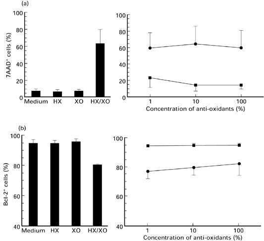
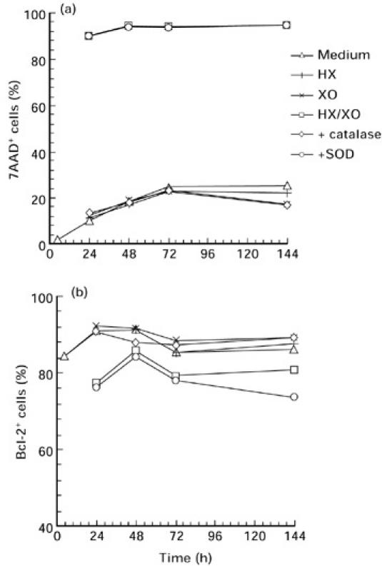
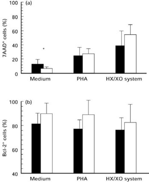
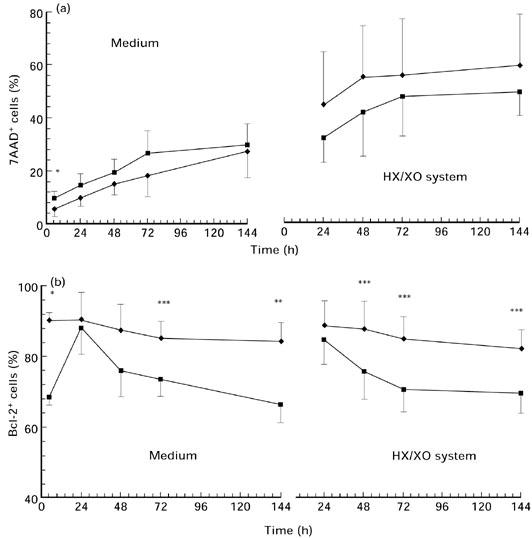
T cell lymphopenia in the peripheral blood lymphocytes (PBL) of patients with AT is mainly caused by a decrease of naive CD45RA+/CD4+ cells followed by a predominance of memory CD45RO+ lymphocytes. To relate these findings to the regulation of programmed cell death, we investigated the activation state and apoptotic level of PBL in 12 patients and healthy controls by flow cytometry. In accordance with previous investigations, the number of naive CD4+/CD45RA+ cells was significantly decreased in patients compared with healthy controls. This disturbed balance of CD45RA and CD45RO was also reflected in higher amounts of activated HLA-DR and CD95 expressing cells, with a concomitant decrease of Bcl-2 protected lymphocytes in the T cell population. With regard to its role in preventing oxidative-induced cell death, we analysed Bcl-2 expression and apoptosis in the presence of oxidative stress. In culture, cells of patients are more susceptible to spontaneous programmed cell death. However, in our stress-inducing system (hypoxanthine/xanthine oxidase system) the number of cells undergoing apoptosis was lower in patients' cell populations compared with controls. In addition, preliminary results suggest that Bcl-2 expression and level of spontaneous apoptosis in patients can be modified by IL-2 and interferon-gamma.


Figures

Fig. 2
Fig. 2
Evaluation of the hypoxanthine/xanthine oxidase (HX/XO) system for the induction of oxidative stress in the lymphoblastoid Jurkat cell line. Stimulation of apoptosis was prepared by various doses of HX and XO after 24 h stimulation and detected by 7-aminoactinomycin D (7AAD; ♦) and propidium iodide (PI; ▪) incorporation. Values represent the means ± s.d. of three separate experiments.
Fig. 1
Fig. 1
Dot plots from a single representative flow cytometric analysis of CD95/CD45RO (upper panels) and Bcl-2/CD45RO (lower panels) double-stained lymphocytes of AT patients compared with a healthy control.
Fig. 3
Fig. 3
Lymphocytes of healthy individuals treated with hypoxanthine (HX)/xanthine oxidase (XO) (1.0 mm/10 mU/ml),HX and XO alone (left). Dose-dependent effect of catalase (▪) and superoxide dismutase (SOD; •) on cells treated with HX (1.0 mm) and XO (10 mU) for 24 h (right). One hundred percent means 1000 U catalase and 10 000 U SOD. Mean values ± s.d. of (a) 7-aminoactinomycin D (7AAD) incorporation (n = 8) and (b) number of Bcl-2-expressing cells (n = 4) detected by flow cytometry.
Fig. 4
Fig. 4
Measurement of (a) 7-aminoactinomycin D (7AAD) incorporation and (b) number of Bcl-2-expressing cells from four healthy individuals by flow cytometry. Lymphocytes were cultured in the presence of hypoxanthine (HX)/xanthine oxidase (XO) (1.0 mm/10 mU/ml), HX and XO alone over a period of 144 h. With regard to the inhibitory effect of the radical scavengers catalase (100 U/ml) or superoxide dismutase (SOD) (1000 U/ml) were added to cell cultures treated with the HX/XO system. Four healthy controls with a strong sensitivity to oxidative stress were examined. Results are shown as mean values.
Fig. 5
Fig. 5
Apoptotic measurement after 24-h stimulation with medium alone, phytohaemagglutinin (PHA) 1 μg/ml and hypoxanthine (HX) 1 mm/xanthine oxidase (XO) 10 mU of AT lymphocytes (▪) in comparison with healthy controls (□). Mean values ± s.d. of the (a) 7-aminoactinomycin D (7AAD) incorporation and (b) the number of Bcl-2-expressing cells were detected by flow cytometry; *P < 0.05.
Fig. 6
Fig. 6
Time kinetics of a 144-h stimulation with hypoxanthine (HX) 1 mm/xanthine oxidase (XO) 10 mU and medium alone. 7-aminoactinomycin D (7AAD) incorporation (a) and Bcl-2 expression (b) were detected after stimulation of AT lymphocytes (▪) and control cells (♦) by flow cytometry. Values are shown as mean ± s.d.; *P < 0.001; **P < 0.01; ***P < 0.05.

Similar articles

Cited by

References

    1. Gatti RA, Boder E, Vinters HV, Sparkes RS, Norman A, Lange K. Ataxia-telangiectasia: an interdisciplinary approach to pathogenesis. Medicine. 1991;70:99–117. - PubMed
    1. Lavin MF, Shiloh Y. Ataxia-telangiectasia: a multifaceted genetic disorder associated with defective signal transduction. Curr Opin Immunol. 1996;8:459–64. - PubMed
    1. Shiloh Y, Rotman G. Ataxia-telangiectasia and the ATM gene: linking neurodegeneration, immunodeficiency, and cancer to cell cycle checkpoints. J Clin Immunol. 1996;16:254–60. - PubMed
    1. Paganelli R, Scala E, Scarselli E, Ortolani C, Carmini D, Aiuti F, Fiorelli M. Selective deficiency of CD4+/CD45RA+ lymphocytes in patients with ataxia telangiectasia. J Clin Immunol. 1992;12:84–91. - PubMed
    1. Paganelli R, Capobianchi MR, Ensoli B, D'offizi GP, Facchini J, Dianzani F, Aiuti F. Evidence that defective gamma interferon production in patients with primary immunodeficiencies is due to intrinsic incompetence of lymphocytes. Clin Exp Immunol. 1988;72:124–9. - PMC - PubMed

MeSH terms