

Changing myoepithelial cell distribution during regeneration of rat parotid glands
- PMID: 10607019
- PMCID: PMC2517833
- DOI: 10.1046/j.1365-2613.1999.00124.x
Changing myoepithelial cell distribution during regeneration of rat parotid glands
Abstract
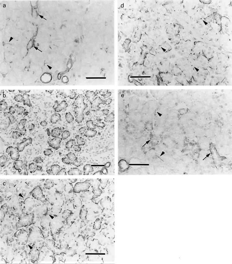
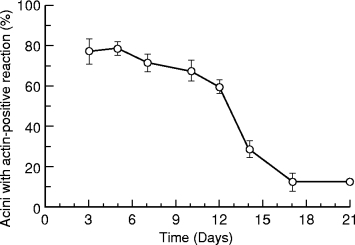
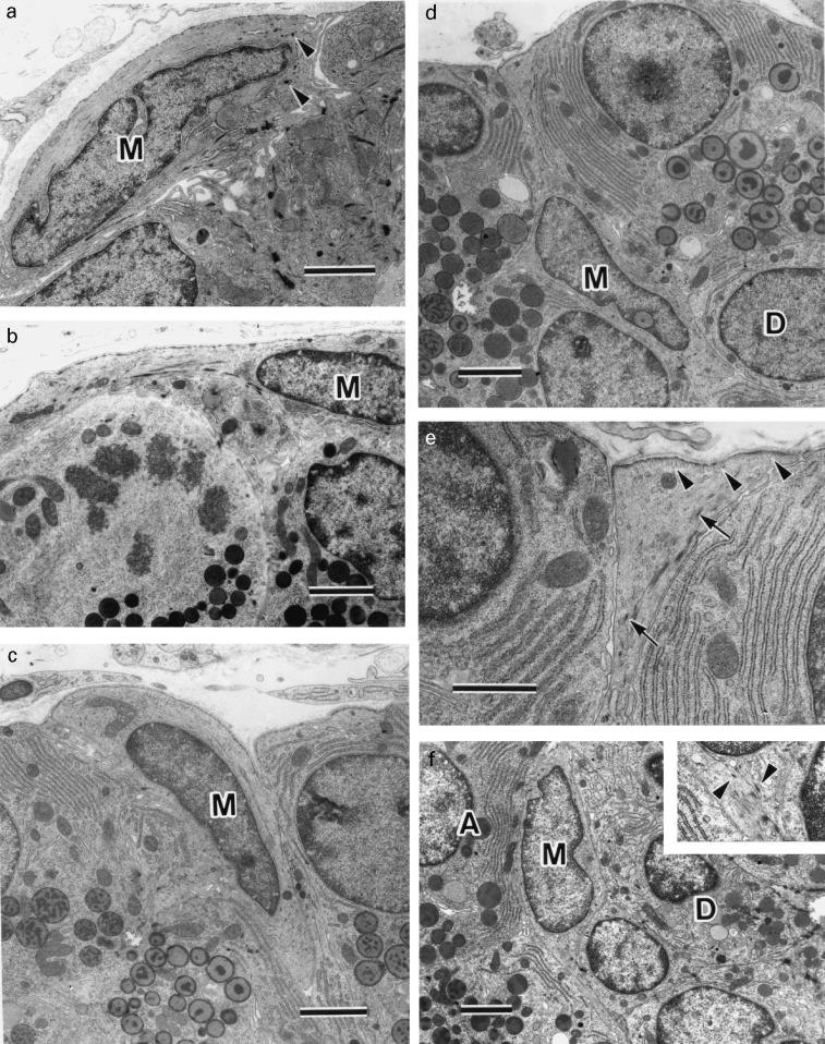
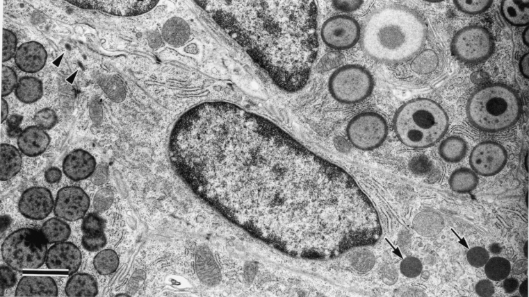
The distribution of the myoepithelial cells during regeneration of the rat parotid gland after atrophy induced by one week of parotid duct ligation was investigated by immunohistochemistry for actin and transmission electron microscopy (TEM). Immunohistochemically, residual ducts were surrounded by actin-positive cells when clips were removed from the duct. Three days later, most of the newly formed acini originating from the residual ducts were also embraced by actin-positive cells. After 10 days, actin-positivity tended to be seen as dots around acini that decreased in number day by day. On day 21 actin-positive cells mainly surrounded intercalated ducts with only a few positive reactions identified at the acinar periphery. Electron microscopically, residual ducts and newly formed acini were peripherally embraced by myoepithelial cells before day 5. After day 7, shift of myoepithelial cells from the periphery of acini to the duct-acinar junctional region was identified. Then few myoepithelial cells were identified at the periphery of acini. These observations indicate that myoepithelial cells migrate from the acinar periphery to the duct-acinar junctional region during rat parotid regeneration, and that such behaviour is closely related to that seen during rat parotid development.
Figures
Similar articles
-
Mitotic proliferation of myoepithelial cells during regeneration of atrophied rat submandibular glands after duct ligation.J Oral Pathol Med. 2004 Aug;33(7):430-4. doi: 10.1111/j.1600-0714.2004.00234.x. J Oral Pathol Med. 2004. PMID: 15250836
-
Changes in the number and distribution of myoepithelial cells in the rat parotid gland during postnatal development.Anat Embryol (Berl). 2006 Oct;211(5):567-74. doi: 10.1007/s00429-006-0111-3. Epub 2006 Aug 26. Anat Embryol (Berl). 2006. PMID: 16937148
-
Biological behavior of myoepithelial cells in the regeneration of rat atrophied sublingual glands following release from duct ligation.J Mol Histol. 2005 Jun;36(5):373-9. doi: 10.1007/s10735-005-9009-2. Epub 2005 Nov 9. J Mol Histol. 2005. PMID: 16283425
-
Functional morphology of myoepithelial cells in the rat salivary glands: A review.J Oral Biosci. 2025 Mar;67(1):100592. doi: 10.1016/j.job.2024.100592. Epub 2024 Nov 29. J Oral Biosci. 2025. PMID: 39615670 Review.
-
Myoepithelial cells in pathology.J Pharm Bioallied Sci. 2015 Apr;7(Suppl 1):S190-3. doi: 10.4103/0975-7406.155898. J Pharm Bioallied Sci. 2015. PMID: 26015706 Free PMC article. Review.
Cited by
-
Experimental Animal Model Systems for Understanding Salivary Secretory Disorders.Int J Mol Sci. 2020 Nov 10;21(22):8423. doi: 10.3390/ijms21228423. Int J Mol Sci. 2020. PMID: 33182571 Free PMC article. Review.
-
Morphological Changes of Myoepithelial Cells in the Rat Submandibular Gland Following the Application of Surgical Stimuli.Acta Histochem Cytochem. 2016 Dec 28;49(6):159-169. doi: 10.1267/ahc.16017. Epub 2016 Dec 23. Acta Histochem Cytochem. 2016. PMID: 28127104 Free PMC article.
-
Early markers of regeneration following ductal ligation in rat submandibular gland.Cell Tissue Res. 2008 May;332(2):227-35. doi: 10.1007/s00441-008-0588-6. Epub 2008 Mar 12. Cell Tissue Res. 2008. PMID: 18335244 Free PMC article.
-
A single injection of interleukin-1 induces reversible aqueous-tear deficiency, lacrimal gland inflammation, and acinar and ductal cell proliferation.Exp Eye Res. 2007 May;84(5):894-904. doi: 10.1016/j.exer.2007.01.015. Epub 2007 Feb 4. Exp Eye Res. 2007. PMID: 17362931 Free PMC article.
-
On approaches to the functional restoration of salivary glands damaged by radiation therapy for head and neck cancer, with a review of related aspects of salivary gland morphology and development.Biotech Histochem. 2008 Jun;83(3-4):103-30. doi: 10.1080/10520290802374683. Biotech Histochem. 2008. PMID: 18828044 Free PMC article. Review.
References
-
- Batsakis JG, Kraemer B, Sciubba JJ. The pathology of head and neck tumors: The myoepithelial cell and its participation in salivary gland neoplasia. Head Neck Surg. 1983;5:222–233. - PubMed
-
- Batsakis JG, Regezi JA, Luna MA, El-Naggar A. Histogenesis of salivary gland neoplasms: a postulate with prognostic implications. J. Laryngol. Otol. 1989;103:939–944. - PubMed
-
- Bhasker SN, Lilly GE, Bhussry B. Regeneration of the salivary glands in the rabbit. J. Dent. Res. 1966;45:37–41. - PubMed
-
- Bogart BI. The fine structural localization of acetylcholinesterase activity in the rat parotid and sublingual glands. Am. J. Anat. 1971;132:259–266. - PubMed
-
- Burford-Mason AP, Cummins MM, Brown DH, MacKay AJ, Dardick I. Immunohistochemical analysis of the proliferative capacity of duct and acinar cells during induced atrophy and subsequent regeneration of rat parotid gland. J. Oral Pathol. Med. 1993;22:440–446. - PubMed
Publication types
MeSH terms
Substances
LinkOut - more resources
Full Text Sources
