

Expression of gelatinase B in trachomatous conjunctivitis
- PMID: 10611105
- PMCID: PMC1723241
- DOI: 10.1136/bjo.84.1.85
Expression of gelatinase B in trachomatous conjunctivitis
Abstract
Background/aims: Gelatinase B is a matrix metalloproteinase involved in extracellular matrix (ECM) breakdown often associated with scarring and other pathological disorders. It was investigated whether gelatinase B is involved in the pathogenesis of ECM degradation associated with trachomatous conjunctivitis.
Methods: Conjunctival biopsy specimens obtained from six patients with active trachoma, six patients with active vernal keratoconjunctivitis (VKC), and seven control subjects were studied. Immunohistochemical techniques and a specific monoclonal antibody against human gelatinase B were used, and a monoclonal antibody against macrophage CD68 to identify mononuclear cells with gelatinase B immunoreactivity. In addition, quantitative zymography was used to compare the activity of gelatinase B in conjunctival biopsy specimens from seven patients with active trachoma and seven control subjects.
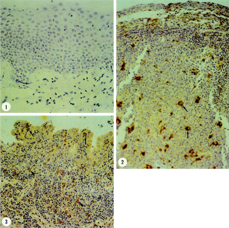
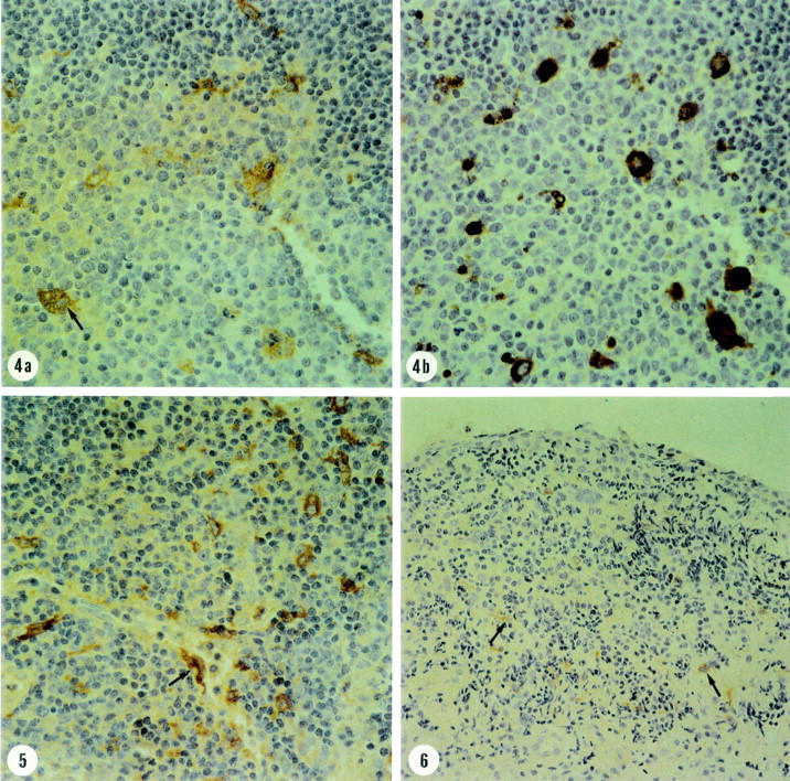
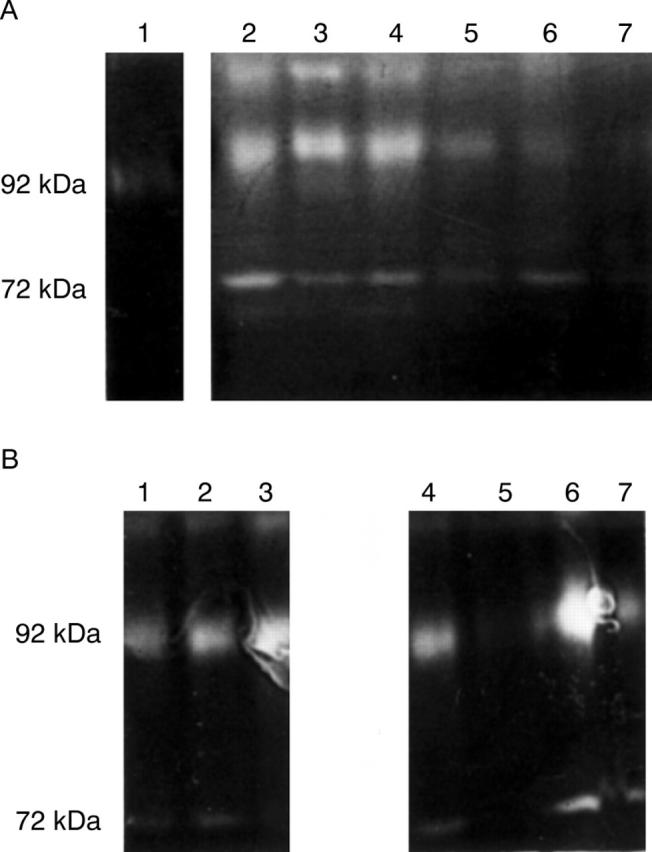
Results: Gelatinase B was detected by immunohistochemistry only in polymorphonuclear cells located in the vascular lumens in three normal conjunctival biopsy specimens. In all trachoma specimens and in five VKC specimens, gelatinase B was localised in monocyte/macrophage cells, positive for the CD68 marker, and in polymorphonuclear cells. The majority of the latter cell type was located in intravascular spaces. Compared with VKC specimens, trachoma specimens showed significantly more immunoreactive gelatinase B monocyte/macrophage cells (52.3 (21.9) v 8.2 (6.4); p <0.001) and polymorphonuclear cells (23.2 (14.2) v 6.3 (5.4); p = 0. 013). Activated macrophages with giant cell morphology clearly stained with the gelatinase B specific monoclonal antibody were observed in trachoma specimens. Zymography revealed that gelatinase B levels in trachoma specimens were significantly higher than the levels found in normal conjunctiva (1739.6 (1078.3) v 609.3 (395.9) scanning units; p = 0.0127).
Conclusions: The increased activity of gelatinase B and numbers of inflammatory cells containing gelatinase B in trachoma specimens suggest that this enzyme plays a part in the pathogenesis of conjunctival scarring in trachoma.
Figures
References
MeSH terms
Substances
LinkOut - more resources
Full Text Sources
Medical