

Sponge delivery variables and tissue levels of 5-fluorouracil
- PMID: 10611106
- PMCID: PMC1723231
- DOI: 10.1136/bjo.84.1.92
Sponge delivery variables and tissue levels of 5-fluorouracil
Abstract
Aim: To study how the delivery of 5-fluorouracil (5-FU) to ocular tissues is affected by altering delivery variables.
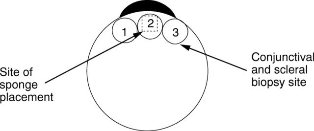
Method: Sponge(s) soaked in radiolabelled 5-FU were placed between the conjunctiva and sclera of pig eyes. Application time, sponge size, sponge make (Altomed, Weck, Merocel), and 5-FU concentration were varied. Conjunctival and scleral tissue levels were determined in samples taken from the application site.
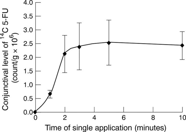
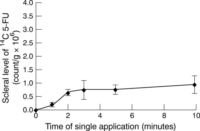
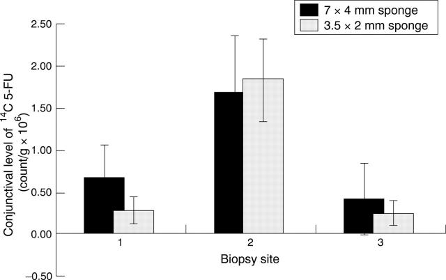
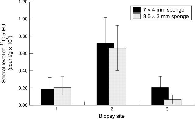
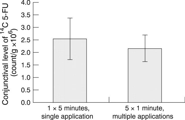
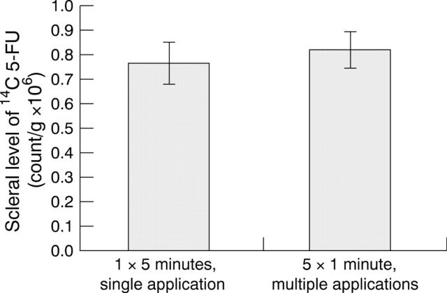
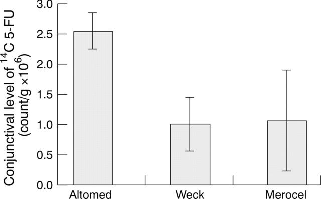
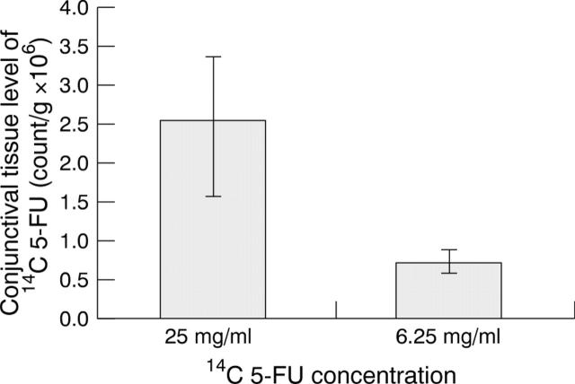
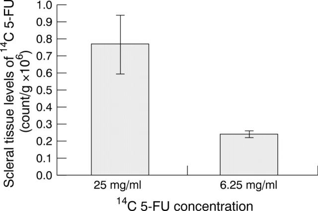
Results: Dose-response curves for scleral and conjunctival 5-FU levels against application time showed increasing tissue levels that reached a plateau after 2-3 minutes. Application beyond 3 minutes did not increase tissue levels. There was no difference in tissue levels between 7x4 and 3. 5x2 mm sponges. Altomed sponges produced 5-FU tissue levels that were twice as high as those obtained with Weck-cell (p<0.01) or Merocel (p<0.02) sponges. Changing the 5-FU concentration from 25 mg/ml to 6.25 mg/ml reduced the conjunctival concentration by a factor of 3.5 (p<0.003).
Conclusion: Application time up to 3 minutes, sponge make, and 5-FU concentration can have a large effect on the tissue delivery of 5-FU. Application time beyond 3 minutes, using 3.5x2 mm or 7x4 mm sponges, and replacing sponges every minute did not have a significant effect on tissue levels. This study models the effect that different variables can have on the ocular tissue levels of an antimetabolite applied intraoperatively.
Figures
References
Publication types
MeSH terms
Substances
LinkOut - more resources
Full Text Sources
Other Literature Sources