. 2000 Jan;20(2):516-22.
doi: 10.1128/MCB.20.2.516-522.2000.

The transmembrane mutation G380R in fibroblast growth factor receptor 3 uncouples ligand-mediated receptor activation from down-regulation

Affiliations

The transmembrane mutation G380R in fibroblast growth factor receptor 3 uncouples ligand-mediated receptor activation from down-regulation

E Monsonego-Ornan et al. Mol Cell Biol. 2000 Jan.

Abstract

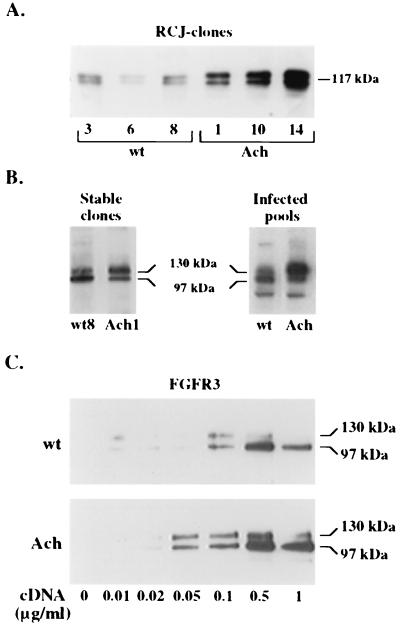
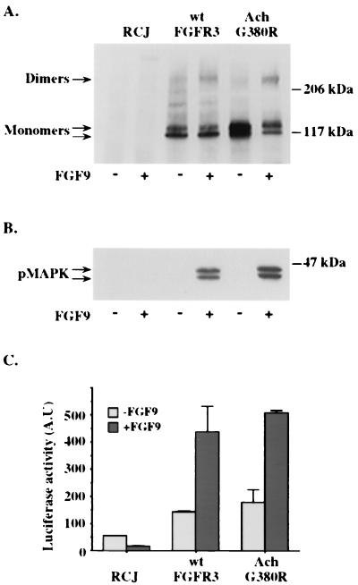
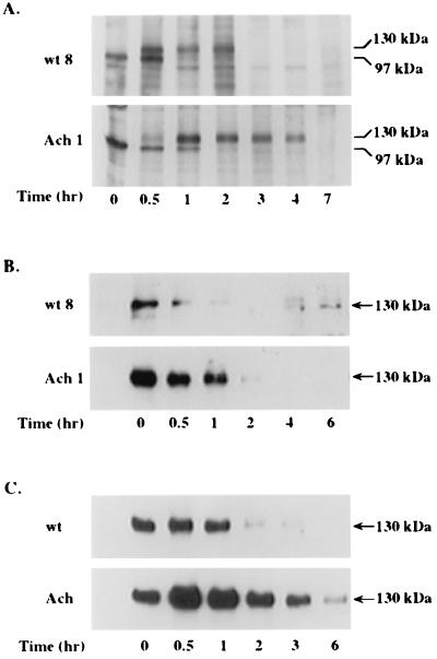
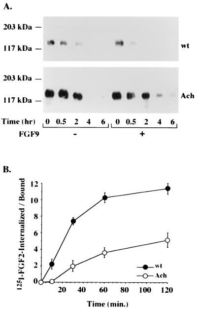
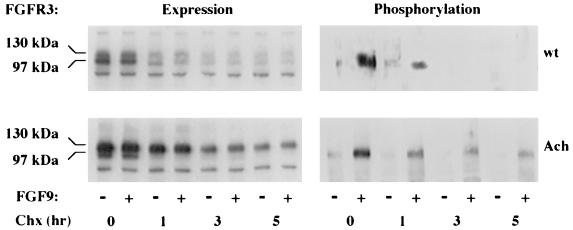
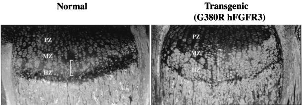
A point mutation, Gly380Arg, in the transmembrane domain of fibroblast growth factor receptor 3 (FGFR3) leads to achondroplasia, the most common form of genetic dwarfism in humans. This substitution was suggested to enhance mutant receptor dimerization, leading to constitutive, ligand-independent activation. We found that dimerization and activation of the G380R mutant receptor are predominantly ligand dependent. However, using both transient and stable transfections, we found significant overexpression only of the mutant receptor protein. Metabolic pulse-chase experiments, cell surface labeling, and kinetics of uptake of radiolabeled ligand demonstrated a selective delay in the down-regulation of the mutant receptor. Moreover, this receptor was now resistant to ligand-mediated internalization, even at saturating ligand concentrations. Finally, transgenic mice expressing the human G380R mutant receptor under the mouse receptor transcriptional control demonstrated a markedly expanded area of FGFR3 immunoreactivity within their epiphyseal growth plates, compatible with an in vivo defect in receptor down-regulation. We propose that the achondroplasia mutation G380R uncouples ligand-mediated receptor activation from down-regulation at a site where the levels and kinetics of FGFR3 signals are crucial for chondrocyte maturation and bone formation.


Figures

FIG. 1
FIG. 1
Expression of wild-type and G380R mutant hFGFR3 in RCJ and 293T cells. (A and B) Lysates of RCJ cells expressing the wt receptor or the G380R mutant receptor were precipitated and probed with anti-FGFR3 antibodies. Three different clones expressing wt or mutant (Ach) FGFR3 (A) or stable clones expressing equal amounts of wt or mutant FGFR3 and RCJ pools infected with wt FGFR3- or mutant FGFR3-encoding retroviruses (B) are shown. (C) 293T cells transfected with the indicated concentrations of either receptor cDNA were lysed, precipitated, and analyzed by SDS-PAGE and Western blotting with anti-FGFR3 antibodies.
FIG. 2
FIG. 2
Receptor dimerization, MAPK activation, and c-fos induction by wt and G380R mutant FGFR3. (A) Stable clones of RCJ cells expressing either the wt receptor or the G380R mutant (Ach) receptor were incubated for 2 h at 4°C in the absence (−) or presence (+) of 50 ng of FGF9 per ml. After chemical cross-linking, cells were lysed, immunoprecipitated with anti-FGFR3 C terminus antibodies, and probed on an immunoblot with a polyclonal antibody to the kinase domain of FGFR3. (B) Cells incubated with (+) or without (−) FGF9 for 9 min at 37°C were lysed, and their lysates were separated by SDS-PAGE and immunoblotted with an anti-pMAPK antibody. (C) Cells transfected with the c-fos luciferase vector were incubated with or without FGF9, lysed, and analyzed for luciferase activity. The data shown represent the means ± standard deviations of duplicate transfections normalized for β-galactosidase activity. A.U, arbitrary units.
FIG. 3
FIG. 3
Metabolic labeling and surface biotinylation of wt and G380R mutant FGFR3 expressed in RCJ cells. (A) Stable clones of RCJ cells expressing wt or G380R mutant (Ach) receptors were pulse-labeled with 35S-methionine for 30 min. At the indicated times, cell lysates were immunoprecipitated with anti-FGFR3 antibodies and resolved by SDS–6% PAGE and autoradiography. (B and C) Stable clones (B) or infected pools (C) of RCJ cells were cell surface biotinylated for 45 min. At the indicated times, cell lysates were precipitated with immobilized avidin, analyzed by SDS–6% PAGE, blotted onto a nitrocellulose membrane, and probed with anti-FGFR3 antibodies. Results represent three independent experiments repeated with different RCJ clones.
FIG. 4
FIG. 4
Ligand-mediated receptor internalization. (A) RCJ cells expressing wt or mutant (Ach) receptors were cell surface biotinylated for 45 min and incubated with (+) or without (−) FGF9 (50 ng/ml) for the indicated times. The cell lysates were precipitated with immobilized avidin, analyzed by SDS–6% PAGE, and probed with anti-FGFR3 antibodies. (B) Cells were incubated for 2 h at 4°C with 125I-FGF2 and then transferred to 37°C for the indicated times. At the end of each incubation, low-affinity heparan sulfate-bound ligand was removed with a high-salt buffer, high-affinity receptor-bound ligand was dissociated under low-pH conditions, and the retained intracellular radioactivity was determined by solubilizing the cells in 100 mM NaOH. The results shown are the averages ± standard deviations of triplicate measurements, calculated as the ratio between the internalized ligand and the receptor-bound ligand. Nonspecific binding was subtracted from all data points.
FIG. 5
FIG. 5
Expression and phosphorylation of wt and G380R mutant FGFR3. RCJ cells expressing either wt or mutant (Ach) receptors were treated for the indicated times with 10 μg of cyclohexamide (Chx) per ml at 37°C. Then, the cells were incubated for 10 min with 50 ng of FGF9 per ml and lysed. Each lysate (1 mg of protein) was immunoprecipitated with an antiphosphotyrosine antibody. Samples were separated by SDS–6% PAGE, transferred to nitrocellulose, and blotted with antibodies to FGFR3.
FIG. 6
FIG. 6
Immunohistochemical analysis of epiphyseal growth plates from normal and transgenic G380R mutant hFGFR3-expressing mice. Immunostaining for FGFR3 was performed on sections of proximal tibia growth plates from 8-day-old normal and transgenic littermates with anti-FGFR3 and fluorescein-conjugated antibodies. Cells expressing FGFR3 are indicated by white brackets. The different zones of the growth plate (PZ, proliferating zone; MZ, maturation zone; MZ, hypertrophic zone) are noted.

References

    1. Basilico C, Moscatelli D. The FGF family of growth factors and oncogenes. Adv Cancer Res. 1992;59:115–165. - PubMed
    1. Bellus G A, McIntosh I, Szabo J, Aylsworth A, Kaitila I, Francomano C A. Hypochondroplasia: molecular analysis of the fibroblast growth factor receptor 3 gene. Ann N Y Acad Sci. 1996;785:182–187. - PubMed
    1. Cancedda R, Descalzi Cancedda F, Castagnola P. Chondrocyte differentiation. Int Rev Cytol. 1995;159:265–358. - PubMed
    1. Coffin J D, Florkiewicz R Z, Neumann J, Mort-Hopkins T, Dorn II G W, Lightfoot P, German R, Howles P N, Kier A, O'Toole B A, et al. Abnormal bone growth and selective translational regulation in basic fibroblast growth factor (FGF-2) transgenic mice. Mol Biol Cell. 1995;6:1861–1873. - PMC - PubMed
    1. Colvin J S, Bohne B A, Harding G W, McEwen D G, Ornitz D M. Skeletal overgrowth and deafness in mice lacking fibroblast growth factor receptor 3. Nat Genet. 1996;12:390–397. - PubMed

Publication types

MeSH terms

Substances

LinkOut - more resources