. 2000 Jan;20(2):604-16.
doi: 10.1128/MCB.20.2.604-616.2000.

A nuclear 3'-5' exonuclease involved in mRNA degradation interacts with Poly(A) polymerase and the hnRNA protein Npl3p

Affiliations

A nuclear 3'-5' exonuclease involved in mRNA degradation interacts with Poly(A) polymerase and the hnRNA protein Npl3p

K T Burkard et al. Mol Cell Biol. 2000 Jan.

Abstract

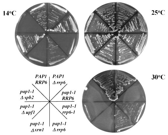
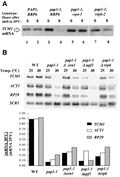
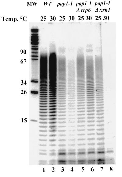
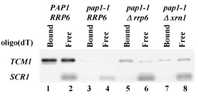
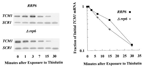
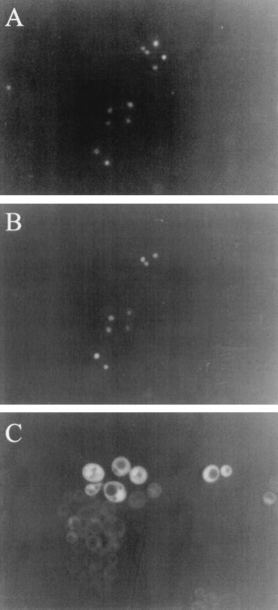
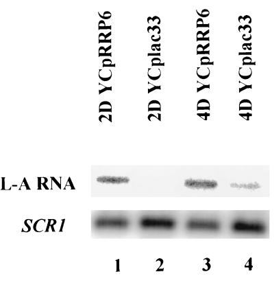
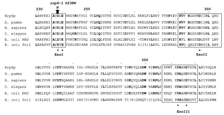
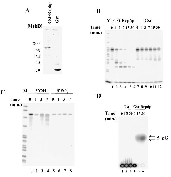
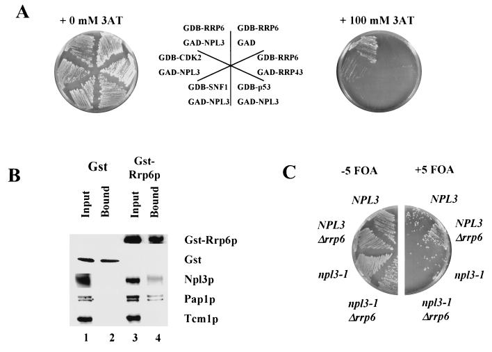
Inactivation of poly(A) polymerase (encoded by PAP1) in Saccharomyces cerevisiae cells carrying the temperature-sensitive, lethal pap1-1 mutation results in reduced levels of poly(A)(+) mRNAs. Genetic selection for suppressors of pap1-1 yielded two recessive, cold-sensitive alleles of the gene RRP6. These suppressors, rrp6-1 and rrp6-2, as well as a deletion of RRP6, allow growth of pap1-1 strains at high temperature and partially restore the levels of poly(A)(+) mRNA in a manner distinct from the cytoplasmic mRNA turnover pathway and without slowing a rate-limiting step in mRNA decay. Subcellular localization of an Rrp6p-green fluorescent protein fusion shows that the enzyme residues in the nucleus. Phylogenetic analysis and the nature of the rrp6-1 mutation suggest the existence of a highly conserved 3'-5' exonuclease core domain within Rrp6p. As predicted, recombinant Rrp6p catalyzes the hydrolysis of a synthetic radiolabeled RNA in a manner consistent with a 3'-5' exonucleolytic mechanism. Genetic and biochemical experiments indicate that Rrp6p interacts with poly(A) polymerase and with Npl3p, a poly(A)(+) mRNA binding protein implicated in pre-mRNA processing and mRNA nuclear export. These findings suggest that Rrp6p may interact with the mRNA polyadenylation system and thereby play a role in a nuclear pathway for the degradation of aberrantly processed precursor mRNAs.


Figures

FIG. 1
FIG. 1
Suppression of pap1-1 temperature sensitivity by mutations in RRP6. Strains BPO2 (PAP1 RRP6), BPO2-12F (PAP1 rrp6::URA3), UR3148-1B (pap1-1), UR3148-1BC-12 (pap1-1 rrp6-1), UR3148-1B-12F (pap1-1 rrp6::URA3), UR3148-1B ΔX (pap1-1 xrn1::URA3), UR3148-1B ΔU (pap1-1 upf1::URA3), and YA201 (pap1-1 spb2::URA3) were grown on YPD plates at the indicated temperatures.
FIG. 2
FIG. 2
Restoration of mRNA levels in pap1-1 strains caused by mutations in RRP6. (A) Steady-state levels of TCM1 mRNA in rrp6 mutants. Total RNA was isolated from strains with the indicated genotypes before and 6 h after a shift to 30°C. TCM1 mRNA was revealed by Northern blot analysis as described in Materials and Methods. (B) Steady-state levels of TCM1, ACT1, and RP29 mRNA and the stable RNA polymerase III transcript SCR1 in pap1-1 mutants. Total RNA was isolated from strains with the indicated genotypes before and 6 h after a shift to 30°C. mRNA levels were revealed by Northern blot analysis as described in Materials and Methods. The bar graphs represent the ratios of mRNA levels after and before a shift to 30°C and were calculated after normalization of the level of each transcript to the level of SCR1 RNA.
FIG. 3
FIG. 3
RRP6 mutation allows the accumulation of poly(A)+ RNA in pap1-1 strains. Total RNA isolated as in Fig. 2 was 3′-end labeled with 5′-[32P]pCp and T4 RNA ligase. After hydrolysis of non-poly(A) tracts, the RNA was separated by PAGE, and the labeled poly(A) was visualized by storage PhosphorImager analysis.
FIG. 4
FIG. 4
RRP6 mutation allows the accumulation of poly(A)+ RNA in pap1-1 strains. Northern blot analysis of total RNA isolated as in Fig. 2 after fractionation on oligo(dT)-cellulose (54). Lanes 1, 3, 5, and 7 contain RNA that was bound to oligo(dT)-cellulose, and lanes 2, 4, 6, and 8 contain RNA that does not bind. Note that RNAs with poly(A) tails of fewer than ca. 20 nucleotides do not bind to oligo(dT) under these conditions; thus, 40 to 50% of yeast mRNAs are not retained on the resin (54).
FIG. 5
FIG. 5
TCM1 mRNA decay rates in RRP6 and Δrrp6 cells. Shown are the results of a Northern blot analysis of TCM1 mRNA levels in total RNA samples from cells as a function of time after treatment with the transcriptional inhibitor thiolutin. The graph on the right illustrates the decay rates of mRNAs from the two strains, plotted after normalization to the levels of the stable SCR1 RNA.
FIG. 6
FIG. 6
Subcellular localization of GFP-Rrp6p in logarithmically growing yeast cells. Strain BPKAN carrying plasmid pGFP-RRP6-FOR11 (A and B) or pGFP-RRP6-REV2 (C) were grown in synthetic complete medium lacking uracil at 30°C to a density of approximately 106/ml. Green fluorescence (B and C) or Hoechst fluorescence (A) was visualized as described in Materials and Methods.
FIG. 7
FIG. 7
RRP6 mutations cause a decrease in LA RNA levels. Northern blot analysis of LA RNA levels from two different rrp6-1 strains carrying the indicated plasmids is shown. LA RNA levels were normalized to SCR1 RNA levels as indicated below.
FIG. 8
FIG. 8
Comparison of the predicted catalytic core of Rrp6p with homologues from Homo sapiens (PM-Scl 100 kDa; Q01780), Schizosaccharomyces pombe (Q10146), C. elegans (P34607), and E. coli (RNase D, P09155; POL, P00582). Amino acid identities occurring in five of the six homologues are highlighted in boldface. ExoI, ExoII, and ExoIII indicate the portions of the sequence homologous to the exonuclease domains of DNA polymerase I and are set off by boxes. The asterisks below the sequences indicate the positions of amino acids essential for the two-metal ligand mechanism of exonuclease activity of DNA polymerase I. The comparison is adapted from reference , which compares a larger set of sequences.
FIG. 9
FIG. 9
Exonuclease activity of recombinant Rrp6p. (A) SDS-PAGE analysis of GST-Rrp6p and GST. After gel electrophoretic separation, the gel was stained with SYBRO RED (Molecular Probes) and analyzed by fluorimager analysis. (B) Storage phosphorimager analysis of PAGE separation of the products of incubation of 5′-32P-labeled CYC1 RNA (10 nM) with GST-Rrp6p (0.1 nM; lanes 1 to 6) or GST (0.1 nM; lanes 7 to 12) for the indicated amounts of time at 30°C. (C) Same as in panel B except that the substrate is 5′-32P-labeled E. coli trpt′ RNA (lanes 3 and 4) or trpt′ RNA 3′-end labeled with 5′-[α-32P]pCp (lanes 5 to 8). (D) Thin-layer chromatographic analysis of the products of incubation of GST (0.5 nM; lanes 1 to 3) or GST-Rrp6p (0.5 nM; lanes 4 to 6) with 5′-[α-32P]GTP-labeled CYC1 RNA (10 nM). Reactions were carried out and analyzed as described in Materials and Methods. The arrow at the right of the figure indicates the position of the 5′pG standard included on the thin-layer chromatography plate.
FIG. 10
FIG. 10
Interaction of Rrp6p with Npl3p and Pap1p. (A) Growth at 30°C of strains carrying various GAL4 DNA binding domain (GBD) fusions and GAL4 activation domain (GAD) fusions on plates with or without the addition of 100 mM 3-AT. The diagram in the center indicates the position of each strain on the adjacent plates. (B) Western blot analysis of proteins bound to glutathione-Sepharose 4B beads from lysates of cells expressing GST (lanes 1 and 2) or GST-Rrp6p (lanes 3 and 4). Input (lanes 1 and 3) represents protein extracts prior to incubation with glutathione-Sepharose 4B beads, and Bound (lanes 2 and 4) represents proteins bound after incubation with glutathione-Sepharose 4B beads (see Materials and Methods). Each of the proteins listed to the right of the figure was detected with specific antisera described in Materials and Methods. (C) Synthetic lethality of the combination of npl3-1 and Δrrp6. PSY1 (NPL3), PSY1.RT (NPL3 Δrrp6), PSY773 (npl3-1), and PSY773.RT (npl3-1 Δrrp6) containing YCpRRP6 were grown at 25°C in the absence (−5 FOA) or the presence (+5 FOA) of 1 g of 5-fluoroorotic acid per liter, which selects for the loss of YCpRRP6, thereby revealing the phenotypes of the chromosomal alleles indicated in the figure.

References

    1. Allmang C, Petfalski E, Podtelejnikov A, Mann M, Tollervey D, Mitchell P. The yeast exosome and human PM-Scl are related complexes of 3′→5′ exonucleases. Genes Dev. 1999;13:2148–2158. - PMC - PubMed
    1. Anderson J S J, Parker R P. The 3′ to 5′ degradation of yeast mRNAs is a general mechanism for mRNA turnover that requires the SKI2 DEVH box protein and 3′ to 5′ exonucleases of the exosome complex. EMBO J. 1998;17:1497–1506. - PMC - PubMed
    1. Atkin A L, Altamura N, Leeds P, Culbertson M R. The majority of yeast UPF1 co-localizes with polyribosomes in the cytoplasm. Mol Biol Cell. 1995;6:611–625. - PMC - PubMed
    1. Bai C, Elledge S J. Gene identification using the yeast two-hybrid system. Methods Enzymol. 1997;283:141–156. - PubMed
    1. Bartel P L, Fields S. Analyzing protein-protein interactions using two-hybrid system. Methods Enzymol. 1995;254:241–263. - PubMed

Publication types

MeSH terms