. 1999 Dec 21;96(26):14694-9.
doi: 10.1073/pnas.96.26.14694.

Activity-based protein profiling: the serine hydrolases

Affiliations

Activity-based protein profiling: the serine hydrolases

Y Liu et al. Proc Natl Acad Sci U S A. .

Abstract

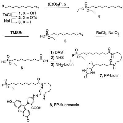
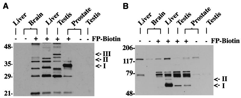
With the postgenome era rapidly approaching, new strategies for the functional analysis of proteins are needed. To date, proteomics efforts have primarily been confined to recording variations in protein level rather than activity. The ability to profile classes of proteins on the basis of changes in their activity would greatly accelerate both the assignment of protein function and the identification of potential pharmaceutical targets. Here, we describe the chemical synthesis and utility of an active-site directed probe for visualizing dynamics in the expression and function of an entire enzyme family, the serine hydrolases. By reacting this probe, a biotinylated fluorophosphonate referred to as FP-biotin, with crude tissue extracts, we quickly and with high sensitivity detect numerous serine hydrolases, many of which display tissue-restricted patterns of expression. Additionally, we show that FP-biotin labels these proteins in an activity-dependent manner that can be followed kinetically, offering a powerful means to monitor dynamics simultaneously in both protein function and expression.


Figures

Scheme 1
Scheme 1
Route for the synthesis of FP-biotin.
Figure 1
Figure 1
FP-biotin reacts with serine hydrolases in an activity-dependent manner. (A) Wild-type FAAH or a mutant FAAH, S241A, in which the enzyme's serine nucleophile was mutated to alanine (80 nM protein) was incubated in either the presence or absence of FP-biotin (2 μM) for 10 min, after which protein was separated from excess inhibitor by SDS/PAGE, electroblotted, and detected by using either avidin or anti-FAAH antibodies. (B) Soluble protein from rat testis (1 μg/μL) was treated with FP-biotin (2 μM) either with or without a preheating step (80°C, 5 min), run on SDS/PAGE (10 μg protein/lane), and visualized by blotting with avidin (Left). Coomassie blue staining confirmed that all lanes contained approximately equal amounts of protein (Right). (C) Rates of reactivity of serine hydrolases with FP-biotin. Testis protein (1 μg/μL) was treated with FP-biotin (2 μM) for the indicated times and analyzed as in B. (Top and Bottom) Taken from film exposures of 1 and 8 min, respectively. (D) (Left) Equal amounts of trypsin (2 μM) were preincubated for 2 hr in either the absence or presence of 1.5 molar equivalents of STI, treated with FP-biotin for 30 min, and analyzed as in B. The lower quantity of trypsin observed in the sample without STI (Top, Coomassie blue staining) is likely the result of a moderate degree of self-proteolysis taking place during the preincubation step. (Right) Testis protein (1 μg/μL) was preincubated with 10 μM STI for 20 min, treated with FP-biotin for 10 min, and analyzed as in B. Arrows highlight proteins whose FP-biotin labeling intensities were reduced significantly (at least 2-fold) in the STI-treated sample relative to a control sample. (Top and Bottom) Taken from film exposures of 1 and 8 min, respectively.
Figure 2
Figure 2
Identification of FP-biotin-reactive proteins from rat brain. (A) Soluble fractions of rat brain (1 μg/μL) were incubated in either the presence or absence of FP-biotin (2 μM) for 30 min and then resolved by SDS/PAGE and blotting with avidin. (B) Separation of rat brain proteins by Q-Sepharose chromatography and labeling of the NaCl elution fractions with FP-biotin. Elution fractions 7–10 (300–500 mM NaCl) are shown. Arrows point to the 75-kDa and 85-kDa FP-biotin-reactive proteins for which sequence information was obtained. (C) The signal intensities of FP-biotin-reactive proteins in fraction 9 were compared with those from a titration of known quantities of purified FAAH reacted to completion with FP-biotin.
Figure 3
Figure 3
FP-biotin detects changes in the expression level of serine hydrolases. (A) Protein samples from HEK-293 cells transfected with a FAAH cDNA, APH cDNA, or empty vector (Mock) were reacted with FP-biotin and resolved by SDS/PAGE (10 μg protein/lane) and blotting with avidin. A strongly labeled 85-kDa protein (I) was detected exclusively in the cytosolic and membrane fractions of APH-transfected cells, whereas a strongly labeled 65-kDa protein (II) was observed specifically in the membrane fractions of FAAH-transfected cells. (B) A longer exposure time of the cytosol blot (2 min vs. 10 sec in A) identified an 85-kDa FP-biotin-reactive protein (I) in the mock and FAAH-transfected HEK cells, possibly representing endogenous levels of APH in this cell type.
Figure 4
Figure 4
FP-biotin identifies several candidate serine hydrolase activities with tissue-restricted patterns of expression. (A) Soluble fractions from indicated rat tissues (1 μg/μL) were treated with FP-biotin (2 μM) and resolved by SDS/PAGE (10 μg protein/lane; 14% polyacrylamide gel) and blotting with avidin. Arrows point to phosphonylated proteins specifically expressed in prostate (I), testis (III), and brain/liver or brain/testis (II). (B) Same as in A, except samples were run on an 8% polyacrylamide gel to more clearly resolve FP-biotin-reactive proteins of higher molecular mass. Arrows point to proteins expressed predominantly (I) or exclusively (II) in liver. Weakly avidin-reactive proteins in the samples untreated with FP-biotin represent putative endogenously biotinylated proteins (18).

Similar articles

Cited by

References

    1. Kalafatis M, Egan J O, van't Veer C, Cawthern K M, Mann K G. Crit Rev Eukaryotic Gene Expression. 1997;7:241–280. - PubMed
    1. Clark J D, Schievella A R, Nalefski E A, Lin L L. J Lipid Mediat Cell Signal. 1995;12:83–117. - PubMed
    1. Mignatti P, Rifkin D B. Enzyme Protein. 1996;49:117–137. - PubMed
    1. Yoshida S, Shiosaka S. Int J Mol Med. 1999;3:405–409. - PubMed
    1. Seidah N G, Chretien M. Curr Opin Biotechnol. 1997;8:602–607. - PubMed

Publication types

MeSH terms

Substances

LinkOut - more resources