Comparative Study
. 1999 Dec 21;96(26):14765-70.
doi: 10.1073/pnas.96.26.14765.

A family of membrane-embedded metalloproteases involved in regulated proteolysis of membrane-associated transcription factors

Affiliations
Comparative Study

A family of membrane-embedded metalloproteases involved in regulated proteolysis of membrane-associated transcription factors

D Z Rudner et al. Proc Natl Acad Sci U S A. .

Abstract

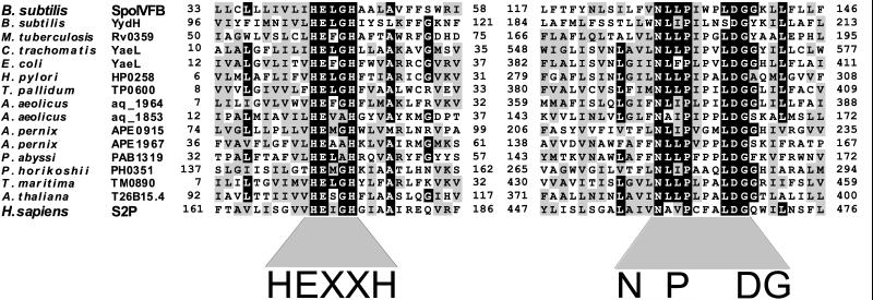
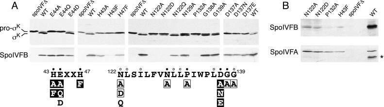
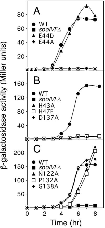
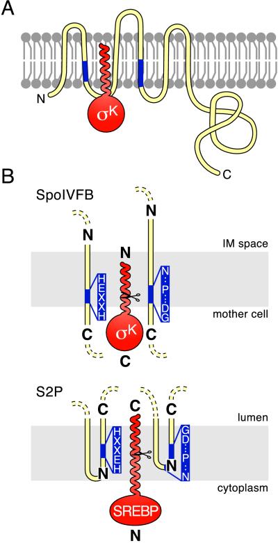
We present evidence that the sporulation protein SpoIVFB of Bacillus subtilis is a member of a newly recognized family of metalloproteases that have catalytic centers adjacent to or within the membrane. SpoIVFB is required for converting the membrane-associated precursor protein, pro-sigma(K), to the mature and active transcription factor sigma(K) by proteolytic removal of an N-terminal extension of 20 amino acids. SpoIVFB and other family members share the conserved sequence HEXXH, a hallmark of metalloproteases, as well as a second conserved motif NPDG, which is unique to the family. Both motifs, which are expected to form the catalytic center of the protease, overlap hydrophobic segments that are predicted to be separate transmembrane domains. The only other characterized member of this family of membrane-embedded metalloproteases is the mammalian Site-2 protease (S2P), which is required for the intramembrane cleavage of the eukaryotic transcription factor sterol regulatory element binding protein (SREBP). We report that amino acid substitutions in the two conserved motifs of SpoIVFB impair pro-sigma(K) processing and sigma(K)-directed gene expression during sporulation. These results and those from a similar analysis of S2P support the interpretation that both proteins are founding members of a family of metalloproteases involved in the activation of membrane-associated transcription factors. Thus, the pathways that govern the activation of the prokaryotic transcription factor pro-sigma(K) and the mammalian transcription factor SREBP not only are analogous but also use processing enzymes with strikingly homologous features.


Figures

Figure 1
Figure 1
Amino acid sequence comparisons of conserved motifs in some members of the newly recognized family of putative metalloproteases. A black box was assigned if 50% of the family members contained the identical residue at that position. A gray box was assigned if 50% of the family members had a similar residue at that position. Similar residues were I, L, V, and M; F, Y, and W; D and E; N and Q; S and T; A and G; and R and K. Amino acid positions are indicated. Gene name and species of origin are shown (Left). An alignment containing the entire 46-member family can be found on our website (http://mcb.harvard.edu/losick).
Figure 2
Figure 2
Pro-σK processing in SpoIVFB mutants. (A) Immunoblots of whole-cell extracts from sporulating cells. All strains analyzed contained a deletion of spoIVF at its normal locus (spoIVFΔ) and a copy of the spoIVF operon at the amyE locus containing either wild-type spoIVFB (WT) or the indicated spoIVFB point mutation. (Upper) Extracts from hour 5 sporulating cells were analyzed by using antibodies that recognize both pro-σK and σK. (Lower) Extracts from hour 3 sporulating cells were analyzed by using antibodies that recognize the C terminus of SpoIVFB (a region that does not contain the two motifs). The results of the analysis are shown schematically below the immunoblots. The amino acid sequences of the two conserved motifs in SpoIVFB are shown in black. Amino acid positions are indicated. Closed and open dots indicate amino acids that are virtually invariant and highly conserved, respectively. Amino acid substitutions that completely abolished pro-σK processing are in black boxes. Mutations that reduced processing efficiency are in black and are surrounded by a box, and those that had no detectable effect on processing are in black. (B) An immunoblot of the four point mutants that had no detectable SpoIVFB protein in A. Twenty-five percent more extract (from hour 3 sporulating cells) was used in this experiment than in those in A. The immunoblot was analyzed by using anti-SpoIVFB antibodies (Upper) and was overexposed to detect the low level of SpoIVFB mutant protein. The immunoblot was reprobed with antibodies that recognize SpoIVFA to control for loading (Lower). The asterisk (*) indicates the position of residual SpoIVFB signal on the reprobed blot.
Figure 3
Figure 3
Analysis of σK-directed β-galactosidase synthesis in SpoIVFB mutant strains. Samples were collected at the indicated times after the initiation of sporulation and assayed for β-galactosidase activity. Each strain contained the gerE-lacZ fusion in the prophage SPβ, a deletion of the spoIVF operon at its normal locus (spoIVFΔ), and a copy of the spoIVF operon at amyE with either wild-type spoIVFB (WT) or a spoIVFB point mutation, as indicated.
Figure 4
Figure 4
Topological models for SpoIVFB, S2P and their substrates. Membranes are shown schematically in gray. Pro-σK and SREBP are in red. SpoIVFB and S2P are in yellow with their conserved motifs in blue (the invariant residues in the two motifs are shown in B). N and C termini are indicated. (A) Proposed insertion of the pro domain of pro-σK into the membrane. Pro-σK is shown with SpoIVFB. The membrane topology of SpoIVFB is based on the work of Green and Cutting (37). The N-terminal pro domain of pro-σK is drawn as a helix based on its predicted secondary structure (40). The mother-cell cytoplasm is below the membrane, and the space between the mother-cell and forespore membranes (the intermembrane space) is above it. (B) Proposed orientation of the catalytic centers in SpoIVFB and S2P. Relevant membrane segments of SpoIVFB and S2P are shown relative to the mother cell or cytoplasmic face of the membrane and to the intermembrane space (IM) or the lumen. Scissors indicate cleavage sites in pro-σK and SREBP. To accommodate the opposite orientation of the transcription factors, the catalytic centers in the two processing enzymes are shown in opposite orientations relative to the compartment into which the mature transcription factors are released (see text).

References

    1. Stragier P, Losick R. Annu Rev Genet. 1996;30:297–241. - PubMed
    1. Brown M S, Goldstein J L. Cell. 1997;89:331–340. - PubMed
    1. Cutting S, Oke V, Driks A, Losick R, Lu S, Kroos L. Cell. 1990;62:239–250. - PubMed
    1. Lu S, Halberg R, Kroos L. Proc Natl Acad Sci USA. 1990;87:9722–9726. - PMC - PubMed
    1. Zhang B, Hofmeister A, Kroos L. J Bacteriol. 1998;180:2434–2441. - PMC - PubMed

Publication types

MeSH terms

LinkOut - more resources