

Defects in transforming growth factor-beta signaling cooperate with a Ras oncogene to cause rapid aneuploidy and malignant transformation of mouse keratinocytes
- PMID: 10611318
- PMCID: PMC24753
- DOI: 10.1073/pnas.96.26.14949
Defects in transforming growth factor-beta signaling cooperate with a Ras oncogene to cause rapid aneuploidy and malignant transformation of mouse keratinocytes
Abstract
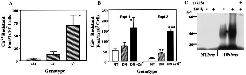
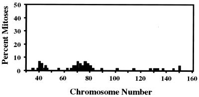
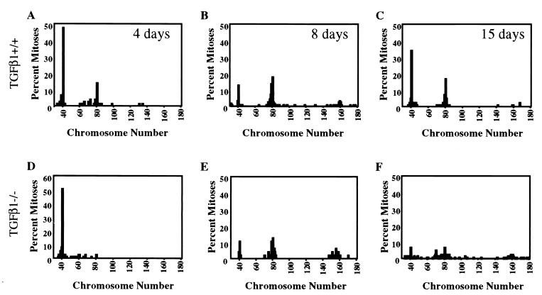
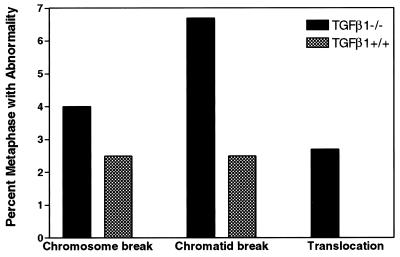
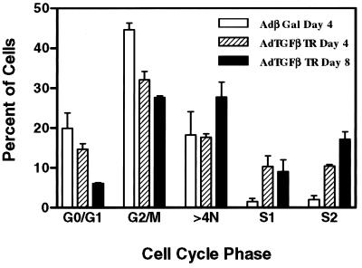
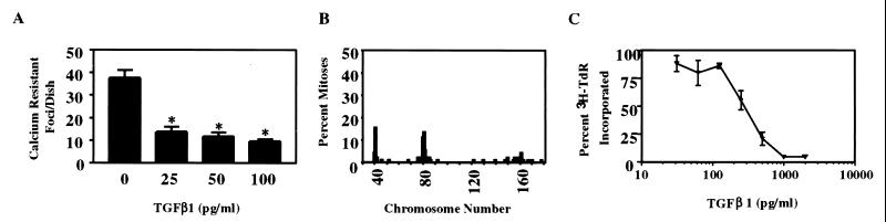
Genetic inactivation of the transforming growth factor-beta (TGF-beta) signaling pathway can accelerate tumor progression in the mouse epidermal model of multistage carcinogenesis. By using an in vitro model of keratinocyte transformation that parallels in vivo malignant conversion to squamous cell carcinoma, we show that v-ras(Ha) transduced primary TGF-beta1-/- keratinocytes and keratinocytes expressing a TGF-beta type II dominant-negative receptor transgene have significantly higher frequencies of spontaneous transformation than control genotypes. Malignant transformation in the TGF-beta1-/- keratinocytes is preceded by aneuploidy and accumulation of chromosomal aberrations. Similarly, transient inactivation of TGF-beta signaling with a type II dominant-negative receptor adenovirus causes rapid changes in ploidy. Exogenous TGF-beta1 can suppress aneuploidy, chromosome breaks, and malignant transformation of the TGF-beta1-/- keratinocytes at concentrations that do not significantly arrest cell proliferation. These results point to genomic instability as a mechanism by which defects in TGF-beta signaling could accelerate tumor progression in mouse multistage carcinogenesis.
Figures
Similar articles
-
Smad7 but not Smad6 cooperates with oncogenic ras to cause malignant conversion in a mouse model for squamous cell carcinoma.Cancer Res. 2003 Nov 15;63(22):7760-8. Cancer Res. 2003. PMID: 14633701
-
Progressive abrogation of TGF-beta 1 and EGF growth control is associated with tumour progression in ras-transfected human keratinocytes.Int J Cancer. 1992 Sep 30;52(3):461-70. doi: 10.1002/ijc.2910520322. Int J Cancer. 1992. PMID: 1328069
-
Restored expression of transforming growth factor beta type II receptor in k-ras-transformed thyroid cells, TGF beta-resistant, reverts their malignant phenotype.J Cell Physiol. 1997 Aug;172(2):200-8. doi: 10.1002/(SICI)1097-4652(199708)172:2<200::AID-JCP7>3.0.CO;2-S. J Cell Physiol. 1997. PMID: 9258341
-
Role of oncogenes and tumor suppressor genes in multistage carcinogenesis.J Invest Dermatol. 1994 Nov;103(5 Suppl):90S-95S. doi: 10.1111/1523-1747.ep12399255. J Invest Dermatol. 1994. PMID: 7963691 Review.
-
TGF-betas and TGF-beta receptors in atherosclerosis.Cytokine Growth Factor Rev. 2000 Mar-Jun;11(1-2):103-14. doi: 10.1016/s1359-6101(99)00034-9. Cytokine Growth Factor Rev. 2000. PMID: 10708958 Review.
Cited by
-
Alcohol, stem cells and cancer.Genes Cancer. 2017 Sep;8(9-10):695-700. doi: 10.18632/genesandcancer.156. Genes Cancer. 2017. PMID: 29234487 Free PMC article. Review.
-
Loss of the transforming growth factor-β effector β2-Spectrin promotes genomic instability.Hepatology. 2017 Feb;65(2):678-693. doi: 10.1002/hep.28927. Epub 2016 Dec 24. Hepatology. 2017. PMID: 28114741 Free PMC article.
-
It takes a tissue to make a tumor: epigenetics, cancer and the microenvironment.J Mammary Gland Biol Neoplasia. 2001 Apr;6(2):213-21. doi: 10.1023/a:1011317009329. J Mammary Gland Biol Neoplasia. 2001. PMID: 11501581 Review.
-
TGF-beta receptor inactivation and mutant Kras induce intestinal neoplasms in mice via a beta-catenin-independent pathway.Gastroenterology. 2009 May;136(5):1680-8.e7. doi: 10.1053/j.gastro.2009.01.066. Epub 2009 Feb 4. Gastroenterology. 2009. PMID: 19208363 Free PMC article.
-
β2-spectrin depletion impairs DNA damage repair.Oncotarget. 2016 Jun 7;7(23):33557-70. doi: 10.18632/oncotarget.9677. Oncotarget. 2016. PMID: 27248179 Free PMC article.
References
-
- Markowitz S, Wang J, Myeroff L, Parsons R, Sun L, Lutterbaugh J, Fan R S, Zborowska E, Kinzler K W, Vogelstein B, et al. Science. 1995;268:1336–1338. - PubMed
-
- Hahn S A, Schutte M, Hoque A T, Moskaluk C A, da Costa L T, Rozenblum E, Weinstein C L, Fischer A, Yeo C J, Hruban R H, et al. Science. 1996;271:350–353. - PubMed
MeSH terms
Substances
Grants and funding
LinkOut - more resources
Full Text Sources
Molecular Biology Databases
