Comparative Study
. 1999 Dec 21;96(26):14955-60.
doi: 10.1073/pnas.96.26.14955.

Modulation of Ca(2+) entry by polypeptides of the inositol 1,4, 5-trisphosphate receptor (IP3R) that bind transient receptor potential (TRP): evidence for roles of TRP and IP3R in store depletion-activated Ca(2+) entry

Affiliations
Comparative Study

Modulation of Ca(2+) entry by polypeptides of the inositol 1,4, 5-trisphosphate receptor (IP3R) that bind transient receptor potential (TRP): evidence for roles of TRP and IP3R in store depletion-activated Ca(2+) entry

G Boulay et al. Proc Natl Acad Sci U S A. .

Abstract

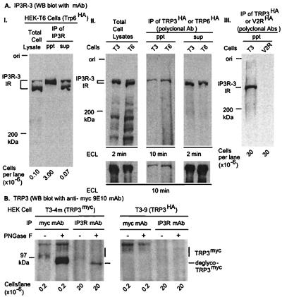
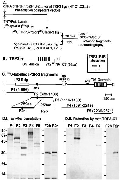
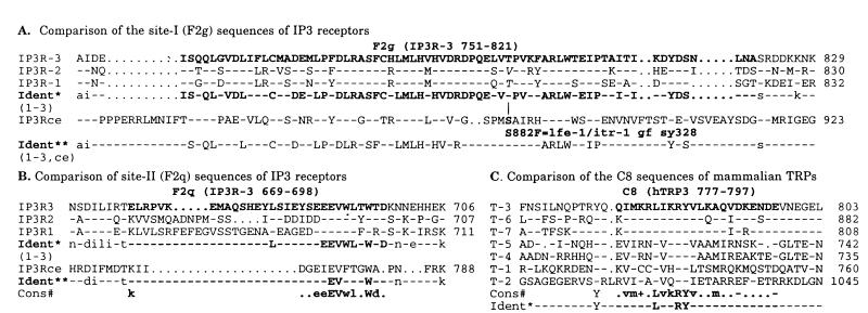
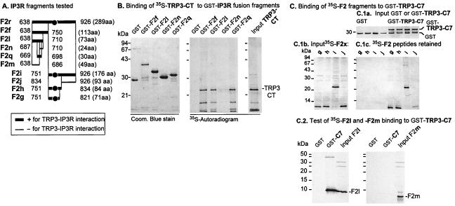
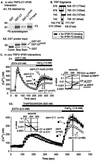
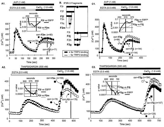
Homologues of Drosophilia transient receptor potential (TRP) have been proposed to be unitary subunits of plasma membrane ion channels that are activated as a consequence of active or passive depletion of Ca(2+) stores. In agreement with this hypothesis, cells expressing TRPs display novel Ca(2+)-permeable cation channels that can be activated by the inositol 1,4,5-trisphosphate receptor (IP3R) protein. Expression of TRPs alters cells in many ways, including up-regulation of IP3Rs not coded for by TRP genes, and proof that TRP forms channels of these and other cells is still missing. Here, we document physical interaction of TRP and IP3R by coimmunoprecipitation and glutathione S-transferase-pulldown experiments and identify two regions of IP3R, F2q and F2g, that interact with one region of TRP, C7. These interacting regions were expressed in cells with an unmodified complement of TRPs and IP3Rs to study their effect on agonist- as well as store depletion-induced Ca(2+) entry and to test for a role of their respective binding partners in Ca(2+) entry. C7 and an F2q-containing fragment of IP3R decreased both forms of Ca(2+) entry. In contrast, F2g enhanced the two forms of Ca(2+) entry. We conclude that store depletion-activated Ca(2+) entry occurs through channels that have TRPs as one of their normal structural components, and that these channels are directly activated by IP3Rs. IP3Rs, therefore, have the dual role of releasing Ca(2+) from stores and activating Ca(2+) influx in response to either increasing IP3 or decreasing luminal Ca(2+).


Figures

Figure 1
Figure 1
Reciprocal coimmunoprecipitation of IP3R and TRP. T3–9 or T6–12 cells, stably expressing HA epitope-tagged TRP3 or TRP6, were lysed, and immunoreactive IP3R was analyzed by Western blotting. (A) IPs of TRP3 and TRP6 contain type-3 IP3R. (I) IP3R-3 in cell lysates, immunoprecipitates (IP, ppt) and IP supernatant (IP, sup). (II) IP3R-3 in detergent extracts and in the respective IP ppt and IP sup. Lower panels of A.II provide an idea of the relative proportion of total IP3R coimmunoprecipitated with TRP3 or TRP6. (III) Lack of significant nonspecific coimmunoprecipitation of IP3R3 immunoreactive material in an IP of V2R, an unrelated integral membrane protein expressed stably in HEK cells (32). Western blot with anti-human IP3R-3 mAb. (B) IPs of IP3R from T3 cell extracts contain TRP3. Detergent extracts of HEK-T3 cells stably expressing TRP6myc or TRP3HA were treated or not with PNGase-F and incubated overnight either with anti-type-1/2/3 IP3R mAb or with 9E10 anti-myc mAb, both preadsorbed to protein-A Sepharose. IPs were washed, dissolved, and electrophoresed as described in Materials and Methods. TRP3myc in IP3R and TRP3 IPs was visualized with 9E10 mAb and detected by enhanced chemiluminescence (ECL).
Figure 2
Figure 2
Binding of IP3 receptor fragments to human TRP3. In vitro-translated IP3R3 F1–F5 fragments, labeled with [35S]Met and [35S]Cys, were incubated with a TRP segment fused to GST and adsorbed to glutathione-Sepharose. After 30 min at room temperature, Sepharose beads were washed, and the adsorbed complexes were resolved by SDS/PAGE, stained, and autoradiographed. In other experiments, the inverse strategy was used: the in vitro-translated fragment was from hTRP3, and the protein fused to GST was from the human IP3R3. (A) Design of the GST-pulldown experiments. (B) Linear diagram of the IP3R3 and TRP3, represented with the same scale and location of some of the molecular landmarks of IP3R3. (C) Representative results showing binding of F2, but not the other IP3R fragments, to TRP-C7.
Figure 3
Figure 3
F2g (A) and F2q (B) IP3R-3 sequences and the C8 TRP sequences (C) are in bold. (A and B) IP3R1, IP3R2, and ceIP3R sequences are compared to that of the IP3R3 sequence (Top). (C) TRP1–TRP7 sequences are compared to that of TRP3 (Top). For these comparisons: - denotes identity, otherwise the amino acid is given in single-letter code. Ident and Cons, identical and consensus amino acids. “Ident,” Identities in upper case. *, Includes the three human IP3Rs. **, Includes three human and one Caenorhabditis elegans IP3R. #, Consensus lists dominant amino acid or amino acid type: +, K or R; –, D or E; ⋅, no consensus. GenBank accession numbers: IP3R1, L38019; IP3R2, D26350; IP3R3, D26351; T-1 (hTRP1), U31110; T-2 (mTRP2), AF111108; T-3 (hTRP3), U47050; T-4 (mTRP4), AF011543; T-5 (mTRP5), AF060107; T-6 (mTRP6), U49069; T-7 (mTRP7), AF139923.
Figure 4
Figure 4
Two regions of IP3R interact with the C-terminus of TRP3. Design and analysis of results is as in Fig. 2. The amino acid compositions of hIP3R3 and hTRP3 correspond to those found in GenBank accession numbers D26351 and U47050, respectively. Beginning and ending amino acid numbers are shown of the fragments translated in vitro or fused to GST.
Figure 5
Figure 5
Binding of wild-type and mutant TRP-C7 to IP3R-F2 and partial interference of agonist and TG-induced Ca2+ entry by TRP3-C7 but not by fragments that do not interact with IP3R. (A) Binding of TRP3 fragments to IP3R-F2. (B) Summary of IP3R-F2 binding to C-terminal fragments of hTRP3. (C) Effect of EBFP and EBFP-TRP fusions on Ca2+ entry stimulated by TG (C2) or by AVP in cells cotransfected with the Gq-coupled V1a receptor (C1). C7, hTRP3[742–795], C7mut3, hTRP3[742–795] with K780D/R781E/K784D/R785E. The strategy and methods described in ref. were used to determine Ca2+ transients. Points are means ± SEM of the number of cells shown analyzed in three to four independent transfections. Insets, Significance of differences between [Ca2+]i curves; P value, Student's t test of the null hypothesis.
Figure 6
Figure 6
Effects of expressing F2q-containing F2l (A) or F2g (C), fused to EBFP, in HEK-293T cells on Ca2+ entry activated either by the Gq-coupled V1a receptor or by TG-induced store depletion. Presentation of data and experimental designs and analyses were the same as in Fig. 5. Points are means ± SEM of the number of cells analyzed in three independent transfections for each of the experiments shown.

Comment in

References

    1. Berridge M J. Biochem J. 1995;312:1–11. - PMC - PubMed
    1. Bird G S J, Putney J W. J Biol Chem. 1993;268:21486–21488. - PubMed
    1. Fasolato C, Innocenti B, Pozzan T. Trends Pharmacol Sci. 1994;15:77–83. - PubMed
    1. Selinger Z, Doza Y N, Minke B. Biochim Biophys Acta. 1993;1179:283–299. - PubMed
    1. Birnbaumer L, Zhu X, Jiang M, Boulay G, Peyton M, Vannier B, Brown D, Platano D, Sadeghi H, Stefani E, Birnbaumer M. Proc Natl Acad Sci USA. 1996;93:15195–15202. - PMC - PubMed

Publication types

Substances