. 1999 Dec 21;96(26):14984-9.
doi: 10.1073/pnas.96.26.14984.

Mice cloned from embryonic stem cells

Affiliations

Mice cloned from embryonic stem cells

T Wakayama et al. Proc Natl Acad Sci U S A. .

Abstract

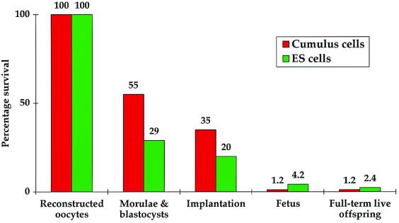
Cloning allows the asexual reproduction of selected individuals such that the offspring have an essentially identical nuclear genome. Cloning by nuclear transfer thus far has been reported only with freshly isolated cells and cells from primary cultures. We previously reported a method of cloning mice from adult somatic cells after nuclear transfer by microinjection. Here, we apply this method to clone mice from widely available, established embryonic stem (ES) cell lines at late passage. With the ES cell line R1, 29% of reconstructed oocytes developed in vitro to the morula/blastocyst stage, and 8% of these embryos developed to live-born pups when transferred to surrogate mothers. We thus cloned 26 mice from R1 cells. Nuclei from the ES cell line E14 also were shown to direct development to term. We present evidence that the nuclei of ES cells at G(1)- or G(2)/M-phases are efficiently able to support full development. Our findings demonstrate that late-passage ES cells can be used to produce viable cloned mice and provide a link between the technologies of ES cells and animal cloning. It thus may be possible to clone from a single cell a large number of individuals over an extended period.


Figures

Figure 1
Figure 1
Selection of E14 cells for subsequent nucleus microinjection into enucleated mouse oocytes. Representatives of small (average diameter 10 μm) and large (arrowhead; average diameter 18 μm) cells were grouped together by using micromanipulators to show contrasting sizes. They are viewed here by Hoffman modulation contrast microscopy at ×200 magnification.
Figure 2
Figure 2
The behavior of nuclear material from small or large ES cells after microinjection into enucleated mouse oocytes. (a) Small cell nuclear components condense to form a disorganized chromosome array 3–4 h after microinjection. (b) Large cell nuclear components condense to form an orderly chromosome array (resembling the maternal metaphase plate) 3–4 h after microinjection. (c) A one-cell embryo formed by the microinjection of a small E14 cell nucleus into an enucleated oocyte and activation by exposure to Sr2+ for 6 h in the presence of cytochalasin B. Two pseudo-pronuclei (pp) are discernible, each containing several nucleoli; remnants of the first polar body (pb1) are also visible. (d) A one-cell embryo formed by the microinjection of a large E14 cell nucleus into an enucleated oocyte and activation by exposure to Sr2+ for 6 h in the absence of cytochalasin B. A degenerate first polar body (pb1), single pseudo-pronucleus (pp), and pseudo-second polar body (indicated by an arrow) are visible.
Figure 3
Figure 3
Mice cloned from ES cells. (a) Hooper (born January 23, 1999) 47 days after birth. He was cloned by using the nucleus of a small E14 cell. He possesses the chinchilla coat color and pink eye phenotypes indicative of the 129/Ola strain. (b) Two cloned mice derived from small R1 ES cells, 9 days after birth. The pups are agouti. The surrogate mother delivered these pups through natural birth.
Figure 4
Figure 4
DNA genotyping corroborates the ES cell provenance of Hooper and other cloned mice. Results for polymorphic DNA markers on chromosome 7 (D7Mit22) and chromosome 4 (D4Mit204) are shown. Confirmation of sex was with PCR primers for the Y-chromosome specific Zfy gene (Zfy). The oocyte donor strain is B6D2F1 (F1), and female surrogates used to carry the pregnancies are of a CD-1 background. (Upper) E14 derived clones. Genomic DNA was from placentae for cloned offspring P1–P6. Genomic DNA from an ear-punch biopsy of Hooper is indicated by H. The weak lower band for the D4Mit204 marker is shared between the placental samples of cloned offspring and the CD-1 sample in a manner consistent with the expected presence of both CD-1-derived (maternal) and clone-derived (E14) cells in the placenta. Accordingly, this lower band is absent from the ear-punch biopsy DNA sample of Hooper (H), whereas it is present in the corresponding placental DNA sample (P1). (Lower) R1-derived clones. Genomic DNA was from tail biopsies for cloned offspring C1–C7.
Figure 5
Figure 5
Survival of cloned embryos at various stages of development. The number of oocytes surviving nuclear transfer is taken as 100%. Progress to the morula/blastocyst stage occurs over 3.5 days in vitro. Sixteen days later, cesarean section is performed, and the number of implantation sites, dead fetuses, and live-born pups are counted. The stage “fetus” includes both dead fetuses and live-born pups.

References

    1. Willadsen S M. Nature (London) 1986;320:63–65. - PubMed
    1. Campbell K H S, McWhir J, Ritchie W A, Wilmut I. Nature (London) 1996;380:64–66. - PubMed
    1. Schnieke A E, Kind A J, Ritchie W A, Mycock K, Scott A R, Ritchie M, Wilmut I, Colman A, Campbell K H. Science. 1997;278:2130–2133. - PubMed
    1. Wilmut I, Schnieke A E, McWhir J, Kind A J, Campbell K H S. Nature (London) 1997;385:810–813. - PubMed
    1. Cibelli J B, Stice S L, Golueke P, Kane J J, Jerry J, Blackwell C, Ponce de Léon F A, Robl J M. Science. 1998;280:1256–1258. - PubMed

Publication types