. 1999 Dec 21;96(26):15062-7.
doi: 10.1073/pnas.96.26.15062.

The human natural killer cell immune synapse

Affiliations

The human natural killer cell immune synapse

D M Davis et al. Proc Natl Acad Sci U S A. .

Abstract

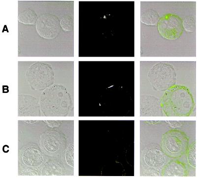
Inhibitory killer Ig-like receptors (KIR) at the surface of natural killer (NK) cells induced clustering of HLA-C at the contacting surface of target cells. In this manner, inhibitory immune synapses were formed as human NK cells surveyed target cells. At target/NK cell synapses, HLA-C/KIR distributed into rings around central patches of intercellular adhesion molecule-1/lymphocyte function-associated antigen-1, the opposite orientation to mature murine T cell-activating synapses. This organization of protein was stable for at least 20 min. Cells could support multiple synapses simultaneously, and clusters of HLA-C moved as NK cells crawled over target cells. Clustering required a divalent metal cation, explaining how metal chelators inhibit KIR function. Surprisingly, however, formation of inhibitory synapses was unaffected by ATP depletion and the cytoskeletal inhibitors, colchicine and cytochalsins B and D. Clearly, supramolecular organization within plasma membranes is critical for NK cell immunosurveillance.


Figures

Figure 1
Figure 1
KIR induce clustering of HLA-C. Various transfectants of 721.221 were imaged when conjugated to peripheral blood NK cells or YTS/KIR1. The first five rows show the Nomarski image of a cell conjugate, the corresponding fluorescence, and an overlay of the fluorescence (colored green for GFP or red for Alexa 594) and Nomarski fields. The target cells are those highlighted by green or red fluorescence. Rows show conjugates of peripheral blood NK line 484 (small cells) and 221/Cw3-GFP (larger cells) (A), YTS/KIR1 and 221/Cw3-GFP (B), YTS/KIR1 and 221/Cw4-GFP (C), YTS/KIR1 and 221/Cw4-GFP in the presence of 50 μg/ml mAb EB6 (anti-KIR1) (D), 221-Cw6, labeled with fluorescent β2m, and YTS/KIR1 (E). (F) YTS/KIR1 and 221/Cw6-GFP, preincubated together, then fixed and permeabilized, stained with 50 μg/ml Alexa-568-conjugated mAb EB6 (red), the corresponding GFP fluorescence (green), and an overlay of the fluorescence fields over the Nomarski image in which colocalized green and red staining is colored yellow. (G) 221/Cw4-GFP conjugated to a peripheral blood NK cell after 0, 10, and 20 min.
Figure 2
Figure 2
Supramolecular organization of the human NK cell immune synapse. Images within the plane of the intercellular contact were taken by laser-scanning confocal microscopy. (A) Distribution of GFP-linked HLA-Cw4 at the contact between 221/Cw4-GFP untransfected both YTS and YTS/KIR1, as well as the intercellular distribution of DiD (a membrane dye) and a mAb to ICAM-1 at the contact between 221/Cw4-GFP and YTS/KIR1. Also shown is the HLA-Cw4/GFP (green) and ICAM-1 (red) distribution overlaid. (B) Intercellular distribution of mAb to LFA-1 or KIR1 on the NK cell (red) in relation to the HLA-C/GFP (green) on the target cell. Superposition of the red and green fluorescence, i.e., where KIR1 and HLA-C staining overlap, is colored yellow. (C) Distribution of HLA-Cw4/GFP and ICAM-1 over 22 min. The diameter of the MHC ring corresponds to the width of MHC clusters at intercellular contacts in Fig. 1.
Figure 3
Figure 3
Clustering of HLA-C by YTS/KIR1 is controlled by metal ions. Rows show conjugates of YTS/KIR1 and 221/Cw4-GFP in the presence of 1 mM 1,10-phenanthroline (A), 1 mM 1,10-phenanthroline and 3 mM ZnCl2 (B), and 1 mM 1,10-phenanthroline and 3 mM MgSO4 (C). For each condition, the Nomarski image of a cell conjugate, the corresponding fluorescence, and an overlay of the fluorescence and Nomarski fields are shown.
Figure 4
Figure 4
The number of NK cell/target cell immune synapses is unaffected by the presence of metabolic or cytoskeletal inhibitors. The first six rows show conjugates of YTS/KIR1 and 221/Cw6-GFP in the presence of 50 mM d-glucose (A), 50 mM azide (B), 13 μM antimycin-A (C), 50 mM 2-deoxyglucose (D), 50 mM 2-deoxyglucose (E), and 10 μM cytochalasin D (NK cell adopts a cone shape in the presence of cytochalasin D) (F). (G) KIR-Ig-coated beads attached to 221/Cw4-GFP. Each row, except E, shows the Nomarski image of a cell conjugate, the corresponding fluorescence, and an overlay of the fluorescence and Nomarski fields. E shows the Nomarski image of a cell conjugate, the corresponding fluorescence image, and the image of the cluster within the plane of the interface (in green box), demonstrating that the ring-like organization of HLA-C also is retained in the absence of ATP.

References

    1. Long E O. Annu Rev Immunol. 1999;17:875–904. - PubMed
    1. Ljunggren H-G, Karre K. Immunol Today. 1990;11:237–244. - PubMed
    1. Colonna M, Brooks E G, Falco M, Ferrera G B, Strominger J L. Science. 1993;260:1121–1124. - PubMed
    1. Colonna M, Borsellino G, Falco M, Ferrera G B, Strominger J L. Proc Natl Acad Sci USA. 1993;90:12000–12004. - PMC - PubMed
    1. Bjorkman P J, Saper M A, Samraoui B, Bennett W S, Strominger J L, Wiley D C. Nature (London) 1987;329:506–512. - PubMed

Publication types

MeSH terms