. 1999 Dec 21;96(26):15074-9.
doi: 10.1073/pnas.96.26.15074.

Elimination of residual metastatic prostate cancer after surgery and adjunctive cytotoxic T lymphocyte-associated antigen 4 (CTLA-4) blockade immunotherapy

Affiliations

Elimination of residual metastatic prostate cancer after surgery and adjunctive cytotoxic T lymphocyte-associated antigen 4 (CTLA-4) blockade immunotherapy

E D Kwon et al. Proc Natl Acad Sci U S A. .

Abstract

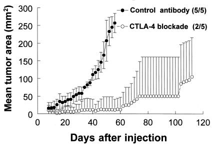
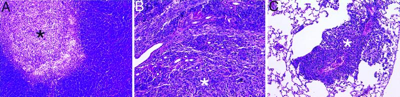
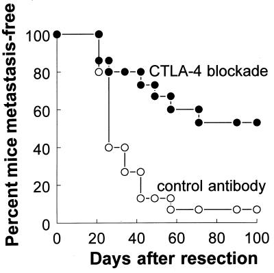
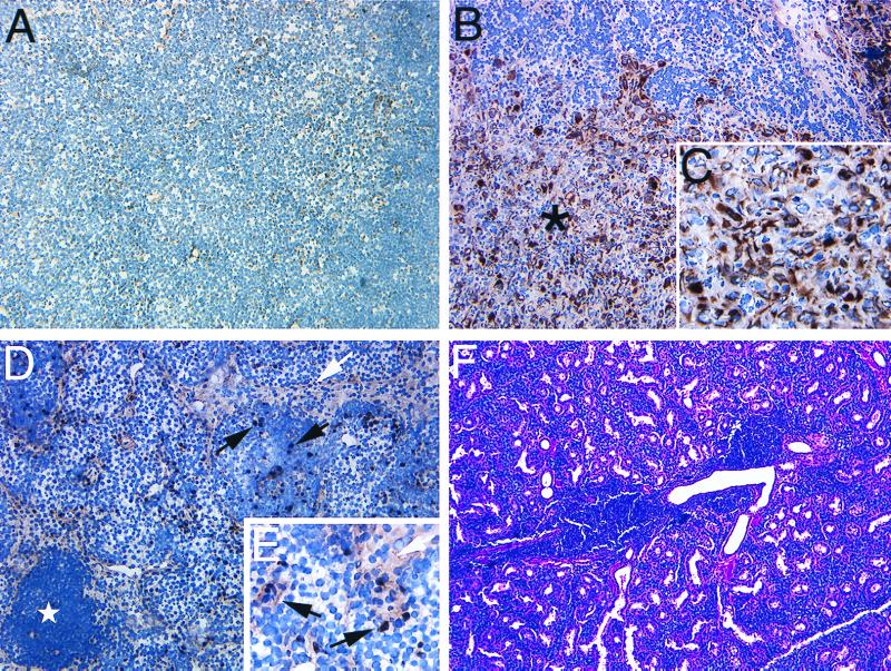
Cancer relapse after surgery is a common occurrence, most frequently resulting from the outgrowth of minimal residual disease in the form of metastases. We examined the effectiveness of cytotoxic T lymphocyte-associated antigen 4 (CTLA-4) blockade as an adjunctive immunotherapy to reduce metastatic relapse after primary prostate tumor resection. For these studies, we developed a murine model in which overt metastatic outgrowth of TRAMP-C2 (C2) prostate cancer ensues after complete primary tumor resection. Metastatic relapse in this model occurs reliably and principally within the draining lymph nodes in close proximity to the primary tumor, arising from established metastases present at the time of surgery. Using this model, we demonstrate that adjunctive CTLA-4 blockade administered immediately after primary tumor resection reduces metastatic relapse from 97.4 to 44%. Consistent with this, lymph nodes obtained 2 weeks after treatment reveal marked destruction or complete elimination of C2 metastases in 60% of mice receiving adjunctive anti-CTLA-4 whereas 100% of control antibody-treated mice demonstrate progressive C2 lymph node replacement. Our study demonstrates the potential of adjunctive CTLA-4 blockade immunotherapy to reduce cancer relapse emanating from minimal residual metastatic disease and may have broader implications for improving the capability of immunotherapy by combining such forms of therapy with other cytoreductive measures including surgery.


Figures

Figure 1
Figure 1
In vivo CTLA-4 blockade slows or prevents the outgrowth of C2 tumors. C57BL/6 mice were injected with 2.5 × 10⁶ C2 cells. Data are mean tumor areas from a single representative of three separate experiments in which groups of five mice received 100-μg intraperitoneal injections of either a control antibody or anti-CTLA-4 (CTLA-4 blockade) on days 7, 10, and 13 after tumor cell injection. The fraction of animals in each group that formed tumors is provided in parentheses next to corresponding marker designations. Note that the lower error bars for CTLA-4 blockade-treated mice have been omitted for clarity.
Figure 2
Figure 2
Histology of C2 metastases stained with H&E. (A) Typical C2 metastasis (*) within a lymph node harvested from a mouse bearing a 250-mm2 C2 primary tumor. C2 infiltration of the serous submandibular salivary gland (B) and metastasis to the lung (C) after overt C2 metastatic lymph node relapse following primary tumor resection and administration of control antibody. All sections are shown at ×20.
Figure 3
Figure 3
Effect of adjunctive CTLA-4 blockade on tumor-free survival of mice after surgical resection of primary C2 tumors. Primary C2 tumors were resected from the dorsal neck of male C57BL/6 mice on day 0 as described in Materials and Methods. Subsequently, cohorts of mice were randomized to receive intraperitoneal injections of 100 μg of either anti-CTLA-4 (CTLA-4 blockade) or control antibody on days 4, 7, and 10 after primary tumor resection (15 mice per treatment group). Beginning on day 21 after surgery, mice were evaluated for the presence of overt (visible) tumors arising from axillary and/or anterior cervical lymph nodes or the primary resection site. Mice not experiencing C2 relapse were followed for a minimum of 150 days after treatment. Data are the percent of mice remaining tumor-free (vertical axis) as a function of the number of days after primary tumor resection (horizontal axis). The experiment shown is a single representative (Experiment 2) of three separate experiments. In this experiment, one mouse in the CTLA-4 blockade-treated cohort died immediately after surgery.
Figure 4
Figure 4
Histology of tissues recovered from mice after sham-surgery, or primary tumor resection followed by adjunctive antibody treatment. (A) Two weeks after sham-surgery and antibody treatment of tumor-naive mice, axillary and anterior cervical lymph nodes in proximity to the surgical site were stained for cytokeratin. Weakly cytokeratin-positive stromal epithelial cells can be observed diffusely distributed throughout the lymph nodes of these mice. (B) Intensely cytokeratin-positive C2 metastases (∗) are observed replacing the normal architecture of lymph nodes taken from mice 2 weeks after C2 primary tumor resection and adjunctive treatment with control-antibody. (C) C2 metastasis, ×40. (D) Regional lymph node recovered from mouse 2 weeks after C2 primary tumor resection and CTLA-4 blockade contains scattered cells of intense or intermediate cytokeratin staining. Marked activation of lymph nodes from these mice is evidenced by the presence of follicle (⋆) formation. Intensely cytokeratin-positive cells (black arrows) appear to represent pyknotic C2 cells whereas the large vacuolated complexes of less-intense cytokeratin staining (white arrows) may represent dying C2 cells, phagocytic cells containing tumor debris, or reactive hyperplasia of stromal elements. (E) Lymph node from mouse treated with primary C2 tumor resection and adjunctive CTLA-4 blockade, ×40. (F) Serous submandibular glands of some mice (5/15) treated with adjunctive CTLA-4 blockade contain single or multiple perivascular infiltrates composed of homogenous mononuclear cells. In contrast, mice treated with adjunctive control antibody demonstrate normal glandular morphology without attendant infiltrate formation. Unless otherwise specified, all sections shown at ×20.

References

    1. Leach D, Krummel M, Allison J P. Science. 1996;271:1734–1736. - PubMed
    1. Kwon E D, Hurwitz A A, Foster B A, Madias C, Feldhaus A L, Greenberg N M, Burg M B, Allison J P. Proc Natl Acad Sci USA. 1997;94:8099–8103. - PMC - PubMed
    1. Foster B A, Gingrich J R, Kwon E D, Madias C, Greenberg N M. Cancer Res. 1997;57:3325–3330. - PubMed
    1. Hurwitz A A, Yu T F-Y, Leach D R, Allison J P. Proc Natl Acad Sci USA. 1998;95:1067–1071. - PMC - PubMed
    1. van Elsas A, Hurwitz A A, Allison J P. J Exp Med. 1999;190:355–366. - PMC - PubMed

Publication types