. 1999 Dec 21;96(26):15127-32.
doi: 10.1073/pnas.96.26.15127.

MEKK1 suppresses oxidative stress-induced apoptosis of embryonic stem cell-derived cardiac myocytes

Affiliations

MEKK1 suppresses oxidative stress-induced apoptosis of embryonic stem cell-derived cardiac myocytes

T Minamino et al. Proc Natl Acad Sci U S A. .

Abstract

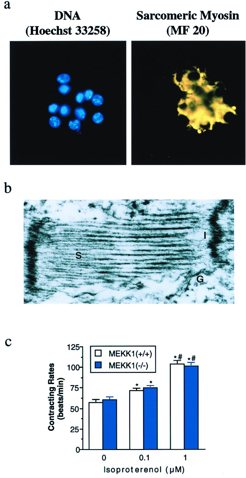
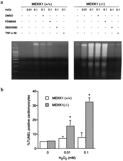
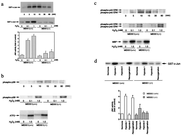
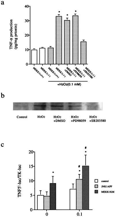
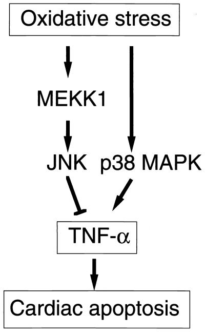
A combination of in vitro embryonic stem (ES) cell differentiation and targeted gene disruption has defined complex regulatory events underlying oxidative stress-induced cardiac apoptosis, a model of postischemic reperfusion injury of myocardium. ES cell-derived cardiac myocytes (ESCM) having targeted disruption of the MEKK1 gene were extremely sensitive, relative to wild-type ESCM, to hydrogen peroxide-induced apoptosis. In response to oxidative stress, MEKK1-/- ESCM failed to activate c-Jun kinase (JNK) but did activate p38 kinase similar to that observed in wild-type ESCM. The increased apoptosis was mediated through enhanced tumor necrosis factor alpha production, a response that was positively and negatively regulated by p38 and the MEKK1-JNK pathway, respectively. Thus, MEKK1 functions in the survival of cardiac myocytes by inhibiting the production of a proapoptotic cytokine. MEKK1 regulation of the JNK pathway is a critical response for the protection against oxidative stress-induced apoptosis in cardiac myocytes.


Figures

Figure 1
Figure 1
Differentiation of MEKK1−/− ES cells into cardiac myocytes. (a) Hoechst 33258 DNA staining of the cluster of MEKK1−/− ESCM after Zeocin selection (Left). Anti-sarcomeric myosin immunofluorescence with MF 20 of the same field (Right). (b) Ultrastructural analysis of selected MEKK1−/− ESCM. S, sarcomere; I, intercalated disc; G, gap junction. (c) Changes in contracting rate of ESCM in response to isoproterenol. Open and solid bars indicate wild-type and MEKK1−/− ESCM (n = 10, each), respectively. * and # indicate significant differences (P < 0.05) from the values at corresponding control and at treatment with 0.1 μM isoproterenol, respectively.
Figure 2
Figure 2
Apoptosis induced by H2O2. (a) H2O2-induced DNA fragmentation in ESCM. Wild-type and MEKK1−/− ESCM were treated with either 0.01 or 0.1 mM H2O2 for 6 h in the presence or absence of either the vehicle (DMSO 0.1%), PD98059 (50 μM), SB 203580 (10 μM), or anti-TNFα antibody (5 μg/ml, purified goat anti-mTNFα IgG; Research and Diagnostic Antibodies, Berkeley, CA). (b) H2O2-induced apoptotic cell death in ESCM evaluated by TUNEL staining. Wild-type and MEKK1−/− ESCM were treated with either 0.01 or 0.1 mM H2O2 for 6 h. Five hundred nuclei of cardiac myocytes costained with Hoechst 33258 were counted and the number of TUNEL positive cells was presented as a percentage from three independent experiments (mean ± SEM). *, P < 0.05 vs. each corresponding control.
Figure 3
Figure 3
MAP kinase activation by oxidative stress. (a) JNK activation by H2O2. (Top) Time course of JNK activation by H2O2 in ESCM. Wild-type ESCM were treated with 1 mM H2O2 for 0–60 min. (Middle and Bottom) Dose-response of JNK activation by H2O2 in wild-type and MEKK1−/− ESCM (15-min treatment). The data represent the average fold of the controls from three independent experiments (mean ± SEM). * and #, P < 0.05 vs. each corresponding control. (b) p38 activation by H2O2. (Top) Phosphorylation of p38 in wild-type ESCM incubated with 1 mM H2O2 for various periods of time. (Middle) Dose response of p38 phosphorylation by H2O2 in wild-type and MEKK1−/− ESCM. (Bottom) Activation of p38 in wild-type and MEKK1−/−ESCM treated with 1 mM H2O2 for 10 min. (c) ERK activation by H2O2. (Top) Phosphorylation of ERK in wild-type ESCM incubated with 1 mM H2O2 for various periods of time. (Middle) Dose response of ERK phosphorylation by H2O2 in wild-type and MEKK1−/− ESCM. (Bottom) Activation of ERK2 in wild-type and MEKK1−/− ESCM treated with 1 mM H2O2 for 10 min. (d) JNK activation by hypoxia/reoxygenation. Wild-type and MEKK1−/− ESCM were subjected to a hypoxic atmosphere by immediately replacing the medium with the hypoxic medium. Cells were incubated in a hypoxic condition for 10 min (Hypoxia-1) or 60 min (Hypoxia-2). After 60-min treatment of cells in hypoxia, cells were reoxygenated by immediately replacing the hypoxic medium with a normoxic medium and incubated in a normoxic condition for an additional 15 min. The data in the graph represent the average percentage of the controls from three independent experiments (mean ± SEM). * and #, P < 0.05 vs. each corresponding control.
Figure 4
Figure 4
TNFα production by H2O2 in ES-derived cardiac myocytes. (a) ELISA assay. Wild-type and MEKK1−/− ESCM were treated with 0.1 mM H2O2 for 6 h in the presence and absence of drugs. TNFα concentration in the supernatant was measured as described in Materials and Methods. (b) Immunoblot. MEKK1−/− ESCM were treated with 0.1 mM H2O2 for 6 h in the presence and absence of drugs. Total cellular proteins were extracted and expression of TNFα in cardiac myocytes was detected by immunoblot analysis by using a specific antibody against TNFα. (c) TNFα promoter reporter assay. pGL3TNF (500 ng) and pRL-TK (50 ng) were cotransfected into wild-type ESCM with 3 μg of either pMEKK1-K1253M, pJNK1-APF, or an empty pCEP4 vector by using a liposome method. Cells were incubated for 24 h and treated with hydrogen peroxide (0.1 mM) for an additional 6 h. Luciferase activities were determined by using a luminometer. TNFα promoter activity was normalized for transfection efficiency based on the cotransfected Renilla luciferase reporter construct.
Figure 5
Figure 5
A proposed model for the role of MEKK1 in cardiac myocytes under oxidative stress.

References

    1. Lucchesi B R. Annu Rev Physiol. 1990;52:561–576. - PubMed
    1. Gottlieb R A, Burleson K O, Kloner R A, Babior B M, Engler R L. J Clin Invest. 1994;94:1621–1628. - PMC - PubMed
    1. Werns S W, Lucchesi B R. Trends Pharmacol Sci. 1990;11:161–166. - PubMed
    1. McCord J M. Fed Proc. 1987;46:2402–2406. - PubMed
    1. Meldrum D R, Dinarello C A, Cleveland J C, Jr, Cain B S, Shames B D, Meng X, Harken A H. Surgery. 1998;124:291–296. - PubMed

Publication types

MeSH terms

Substances

LinkOut - more resources