. 1999 Dec 21;96(26):15143-8.
doi: 10.1073/pnas.96.26.15143.

Plasminogen deficiency leads to impaired remodeling after a toxic injury to the liver

Affiliations

Plasminogen deficiency leads to impaired remodeling after a toxic injury to the liver

J A Bezerra et al. Proc Natl Acad Sci U S A. .

Abstract

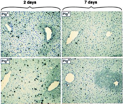
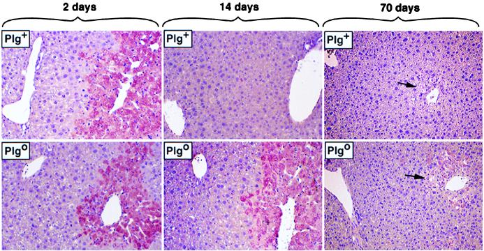
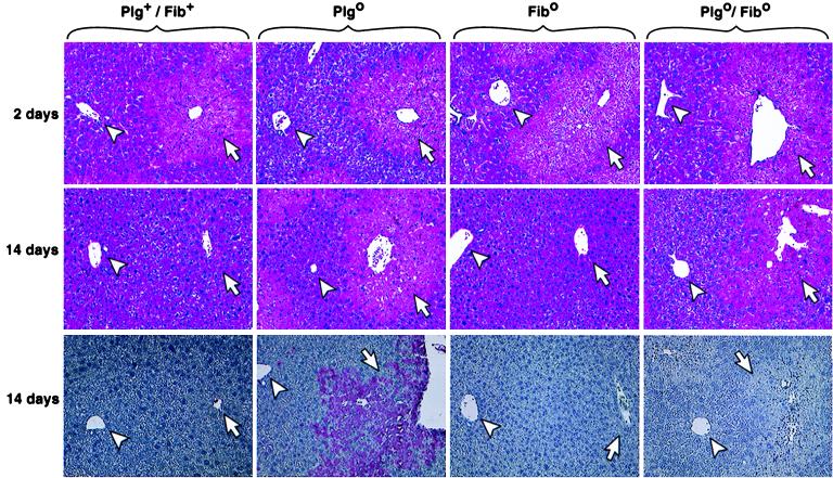
Cellular proliferation and tissue remodeling are central to the regenerative response after a toxic injury to the liver. To explore the role of plasminogen in hepatic tissue remodeling and regeneration, we used carbon tetrachloride to induce an acute liver injury in plasminogen-deficient (Plg(o)) mice and nontransgenic littermates (Plg(+)). On day 2 after CCl(4), livers of Plg(+) and Plg(o) mice had a similar diseased pale/lacy appearance, followed by restoration of normal appearance in Plg(+) livers by day 7. In contrast, Plg(o) livers remained diseased for as long as 2.5 months, with a diffuse pale/lacy appearance and persistent damage to centrilobular hepatocytes. The persistent centrilobular lesions were not a consequence of impaired proliferative response in Plg(o) mice. Notably, fibrin deposition was a prominent feature in diseased centrilobular areas in Plg(o) livers for at least 30 days after injury. Nonetheless, the genetically superimposed loss of the Aalpha fibrinogen chain (Plg(o)/Fib(o) mice) did not correct the abnormal phenotype. These data show that plasminogen deficiency impedes the clearance of necrotic tissue from a diseased hepatic microenvironment and the subsequent reconstitution of normal liver architecture in a fashion that is unrelated to circulating fibrinogen.


Figures

Figure 1
Figure 1
Persistent injury of Plgo livers after CCl4. (Upper) Plg+ livers have a diffuse pale/lacy appearance 2 days after CCl4, followed by restoration of normal appearance by days 7 and 14 (arrows point to residual areas of abnormal appearance). (Lower) In contrast, although the same diseased appearance is seen in Plgo livers on day 2, absence of plasminogen leads to a persistent diseased appearance at days 7 and 14. Although not depicted in the figure, Plgo livers on days 30 and 70 resemble those on day 14.
Figure 2
Figure 2
Persistent centrilobular injury of Plgo livers. Sections of Plg+ livers show an injury to centrilobular hepatocytes 2 days after CCl4, followed by normalization by day 14. In contrast, the centrilobular lesion seen in Plgo livers on day 2 persists at day 14, with no evidence of ongoing repair. At day 70, the centrilobular space of Plgo livers contains a few normal-appearing hepatocytes and abundant dystrophic calcification (Insert), which is also observed sporadically in Plg+ livers (arrows). (Magnifications: panels, ×200; Insert, ×400.)
Figure 3
Figure 3
BrdUrd labeling after liver injury. BrdUrd-hepatocytes are abundant and distributed uniformly throughout the noninjured lobular region 2 days after CCl4 in Plg+ and Plgo livers. Thereafter, labeled hepatocytes are infrequently found in Plg+ livers. Note that labeled hepatocytes are similarly inconspicuous in Plgo livers, despite the persistent centrilobular injury at day 7. (Magnification: ×200.)
Figure 4
Figure 4
Fibrin deposition in Plgo livers. Immunohistochemical staining showing fibrin deposition in diseased areas (pink) of Plg+ and Plgo livers 2 days after CCl4. In Plg+ livers, resolution of injury is accompanied by the timely removal of fibrin. In the absence of plasminogen, fibrin deposits persist up to day 14 after toxic injury; at day 70, minimal residual immunostaining is seen in the area of dystrophic calcification (arrows). (Magnification: ×200.)
Figure 5
Figure 5
Fibrin deficiency does not rescue the lesion of Plgo livers. Liver sections show a similar centrilobular injury regardless of the genotype 2 days after CCl4. On day 14, histology of Plg+/Fib+ and Fibo livers is normal, whereas the lesion persists in Plgo and Plgo/Fibo livers. (Bottom) Immunohistochemical staining shows the characteristic accumulation of fibrin in diseased Plgo livers 14 days after CCl4, whereas no specific staining is noted in diseased Plgo/Fibo livers. (Arrowheads, portal vein; arrows, centrilobular region; magnification: ×200.)

References

    1. Cressman D E, Greenbaum L E, DeAngelis R A, Ciliberto G, Furth E E, Poli V, Taub R. Science. 1996;274:1379–1383. - PubMed
    1. Yamada Y, Kirillova I, Peschon J J, Fausto N. Proc Natl Acad Sci USA. 1997;94:1441–1446. - PMC - PubMed
    1. Rai R M, Lee F Y, Rosen A, Yang S Q, Lin H Z, Koteish A, Liew F Y, Zaragoza C, Lowenstein C, Diehl A M. Proc Natl Acad Sci USA. 1998;95:13829–13834. - PMC - PubMed
    1. Greenbaum L E, Li W, Cressman D E, Peng Y, Ciliberto G, Poli V, Taub R. J Clin Invest. 1998;102:996–1007. - PMC - PubMed
    1. Jakowiew S B, Mead J E, Danielpour D, Wu J, Roberts A B, Fausto N. Cell Regul. 1987;2:535–548. - PMC - PubMed

Publication types

LinkOut - more resources