. 1999 Dec 21;96(26):15233-8.
doi: 10.1073/pnas.96.26.15233.

Apolipoprotein E is essential for amyloid deposition in the APP(V717F) transgenic mouse model of Alzheimer's disease

Affiliations

Apolipoprotein E is essential for amyloid deposition in the APP(V717F) transgenic mouse model of Alzheimer's disease

K R Bales et al. Proc Natl Acad Sci U S A. .

Abstract

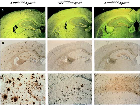
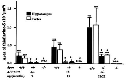
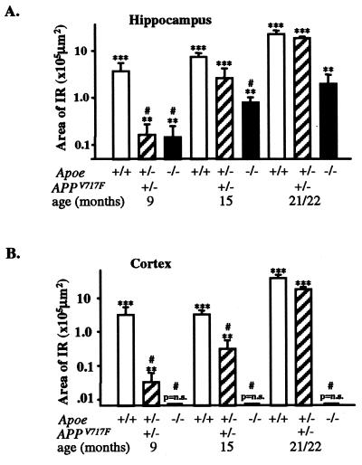
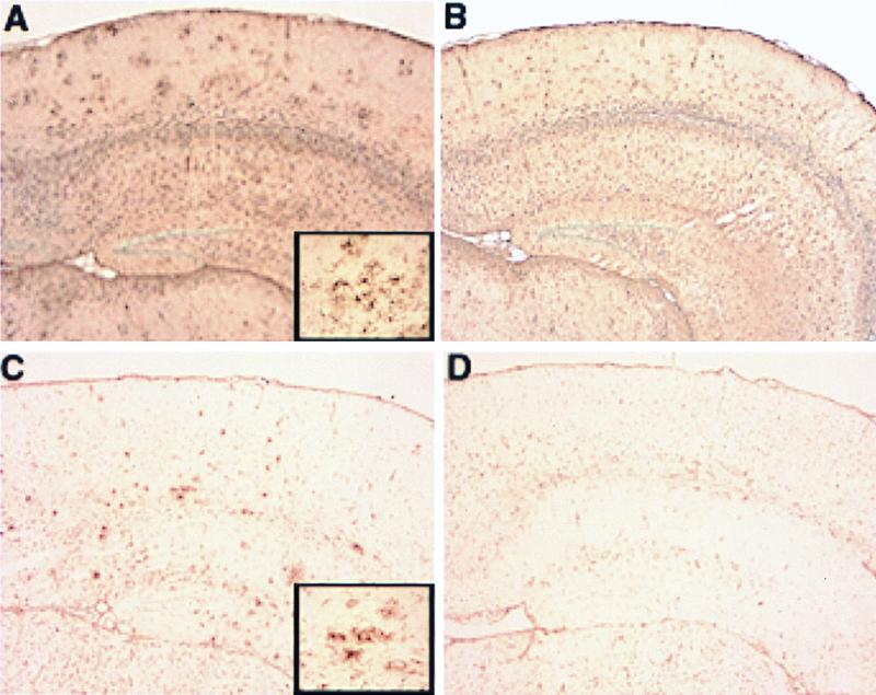
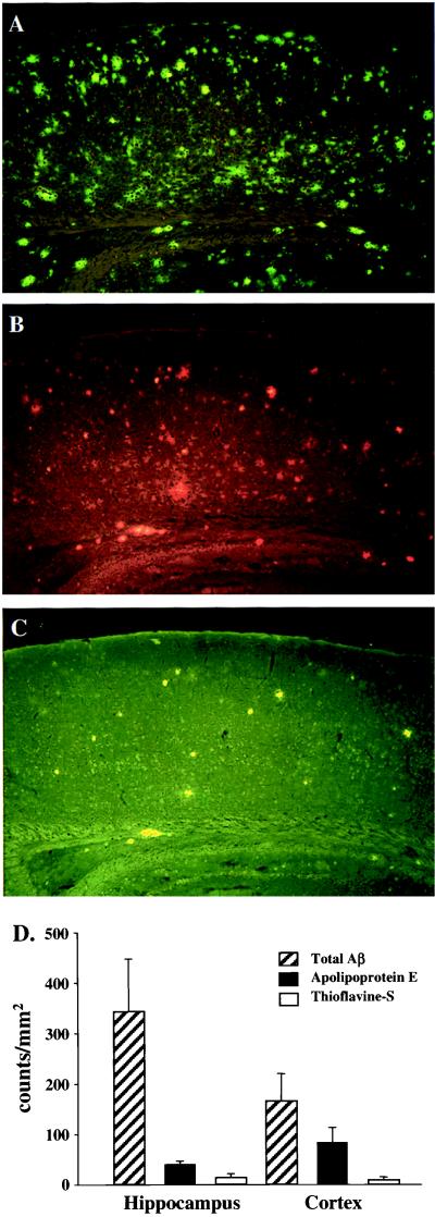
We quantified the amount of amyloid beta-peptide (Abeta) immunoreactivity as well as amyloid deposits in a large cohort of transgenic mice overexpressing the V717F human amyloid precursor protein (APP(V717F+/-) TG mice) with no, one, or two mouse apolipoprotein E (Apoe) alleles at various ages. Remarkably, no amyloid deposits were found in any brain region of APP(V717F+/-) Apoe(-/-) TG mice as old as 22 mo of age, whereas age-matched APP(V717F +/-) Apoe(+/-) and Apoe(+/+) TG mice display abundant amyloid deposition. The amount of Abeta immunoreactivity in the hippocampus was also markedly reduced in an Apoe gene dose-dependent manner (Apoe(+/+) > Apoe(+/-) >> Apoe(-/-)), and no Abeta immunoreactivity was detected in the cerebral cortex of APP(V717F+/-) Apoe(-/-) TG mice at any of the time points examined. The absence of apolipoprotein E protein (apoE) dramatically reduced the amount of both Abeta(1-40) and Abeta(1-42) immunoreactive deposits as well as the resulting astrogliosis and microgliosis normally observed in APP(V717F) TG mice. ApoE immunoreactivity was detected in a subset of Abeta immunoreactive deposits and in virtually all thioflavine-S-fluorescent amyloid deposits. Because the absence of apoE alters neither the transcription or translation of the APP(V717F) transgene nor its processing to Abeta peptide(s), we postulate that apoE promotes both the deposition and fibrillization of Abeta, ultimately affecting clearance of protease-resistant Abeta/apoE aggregates. ApoE appears to play an essential role in amyloid deposition in brain, one of the neuropathological hallmarks of Alzheimer's disease.


Figures

Figure 1
Figure 1
Lack of apoE reduces both Aβ immunoreactive as well as amyloid deposits in a transgenic mouse model of AD. In 21/22-mo-old APPV717+/− TG mice wild-type for Apoe (APPV717F+/− Apoe+/+), numerous thioflavine-S-fluorescent (A) and Aβ immunoreactive deposits (B and C) are evident in both the hippocampus and the cerebral cortex. By contrast, in 21/22-mo-old APPV717F+/− Apoe−/− mice, no thioflavine-S-fluorescent Aβ deposits are observed in any brain region. Furthermore, Aβ-immunoreactive deposits are confined to the hippocampus. APPV717F+/− Apoe+/− mice had an essentially intermediate level of both thioflavine-S-fluorescent and Aβ immunoreactive deposits. Neither thioflavine-S-fluorescent nor Aβ-immunoreactive deposits were found in APPV717F−/− Apoe+/+ mice. The photomicrographs are from representative sections of each of the genotypes indicated. (Original magnification, A and B ×9, C ×35.)
Figure 2
Figure 2
ApoE expression is required for amyloid deposition in the APPV717F TG mouse. APPV717F+/− TG mice with various apoE alleles (Apoe+/+; +/−;−/−) were studied at 9, 15, and 21/22 mo of age. Serial coronal sections were stained with thioflavine-S, and the area occupied by fluorescence was quantified for both the hippocampus and the cerebral cortex (see text for details). Note the complete absence of amyloid deposits in APPV717F+/− TG mice in the absence of Apoe and the modest increase with age in Apoe+/− mice. Amyloid deposition in APPV717F+/− Apoe+/− mice never exceeds 20–30% of that observed in APPV717F+/− Apoe+/+ mice despite significant Aβ immunoreactive deposits in the hippocampus (see Fig. 3). All data were log transformed and tested for normality before statistical analysis (see Materials and Methods). ***, P < 0.001; **, P < 0.01; P = not significant (ns) compared with APPV717F−/− Apoe+/+ mice. #, P < 0.01 compared with APPV717F+/− Apoe+/+ mice.
Figure 3
Figure 3
Effect of apoE on Aβ immunoreactivity in the hippocampus and cerebral cortex of APPV717F+/− TG mice with various Apoe alleles. Serial coronal sections were stained for total Aβ (10D5, 1:1,000), and the area occupied by immunoreactive deposits was quantified (see Materials and Methods). The area occupied by Aβ immunoreactivity increased significantly from 9 to 21/22 mo in APPV717F+/− Apoe+/+ and APPV717F+/− Apoe+/− mice. A reduction in the area occupied by Aβ immunoreactivity was seen in the hippocampus (A) of APPV717F+/− Apoe−/− mice at all ages examined. In contrast to the hippocampus, no Aβ immunoreactive deposits were observed in the cortex (B) of Apoe-deficient APPV717F+/− mice. Note that by 21/22 mo, Apoe hemizygous mice have a similar amount of Aβ immunoreactivity as Apoe homozygous mice, whereas the amount of amyloid deposition is significantly decreased to ≤30% of homozygous mice (Fig. 2). All data were log transformed and tested for normality before statistical analysis (see Materials and Methods). ***, P < 0.001; **, P < 0.01; P = ns compared with APPV717F−/− Apoe+/+ TG mice. #, P < 0.01 compared with APPV717F+/− Apoe+/+ TG mice.
Figure 4
Figure 4
Aβ immunoreactivity in APPV717F+/− TG mice with the three Apoe genotypes: (A) Apoe−/−; (B) Apoe+/−; and (C) Apoe+/+ at 21/22 mo of age. Sections were stained with an antibody specific for Aβ1–42 (21F12; see Materials and Methods for details). There is a decrease in the area occupied by Aβ immunoreactivity in the hippocampus of APPV717F+/− Apoe−/− mice and a complete absence of Aβ immunoreactivity (including total Aβ; see also Fig. 1) in the cerebral cortex (A). The distribution of Aβ1–42 immunoreactivity was similar in all genotypes, with the exception of the dentate gyrus of the hippocampus where the polymorph layer was preferentially affected in APPV717F+/− Apoe−/− mice. Note the absence of Aβ deposits in the outer molecular layer of the dentate gyrus in Apoe−/− compared with Apoe+/+ mice (A vs. B). (Original magnification ×5.)
Figure 5
Figure 5
Reduced glial activation in APPV717F+/− Apoe−/− TG mice. Glial activation was evaluated by GFAP immunohistochemistry (A and B) and tomato lectin histochemistry (C and D) on paraffin-embedded brain sections from 21/22-mo-old APPV717F+/− Apoe+/+ and APPV717F+/− Apoe−/− mice (see Materials and Methods for details). A range of glial staining for each genotype analyzed was apparent with an increase in GFAP immunoreactivity and lectin staining in the superficial layers of the cerebral cortex and hippocampus in APPV717F+/− Apoe+/+ mice (A and C, respectively) compared with APPV717F+/− Apoe−/− mice (B and D, respectively). Note clusters of intensely stained astrocytes (A, Inset) and microglia (C, Inset), which are more prominent in APPV717F+/− Apoe+/+ than in APPV717F+/− Apoe−/− mice. (×5; Inset, ×40.)
Figure 6
Figure 6
ApoE immunoreactivity is associated with a subset of Aβ immunoreactive deposits. (A) Total Aβ immunoreactive deposits (10D5 antibody); (B) apoE immunoreactive deposits; (C) thioflavine-S-fluorescent deposits; and (D) quantitation of Aβ, apoE, and thioflavine-S fluorescent (see Materials and Methods for details). ApoE immunoreactive deposits represent a subpopulation of the total Aβ deposits, but virtually all (≥99%) thioflavine-S-positive Aβ deposits are immunoreactive for apoE. The latter was determined by counting thioflavine-S-fluorescent Aβ deposits in both the hippocampus (n = 273) and cortex (n = 256) and determining by double staining in the same sections (B and C) whether these were also apoE positive (≥99% of thioflavine-S-fluorescent Aβ deposits in each region were apoE positive) (data not shown).

References

    1. Strittmatter W J, Saunders A M, Schmechel D, Pericak-Vance M A, Enghild J, Salvesen G S, Roses A D. Proc Natl Acad Sci USA. 1993;90:1977–1981. - PMC - PubMed
    1. Weisgraber K H, Mahley R W. FASEB J. 1996;10:1485–1494. - PubMed
    1. Corder E H, Saunders A M, Risch N J, Strittmatter W J, Schmechel D E, Gaskell P C, Jr, Rimmler J B, Locke P A, Conneally P M, Schmader K E, et al. Nat Genet. 1994;7:180–184. - PubMed
    1. Lambert J-C, Berr C, Pasquier F, Delacourte A, Frigard B, Cottel D, Pérez-Tur J, Mouroux V, Mohr M, Cécyre D, et al. Hum Mol Genet. 1998;7:1511–1516. - PubMed
    1. Bullido M J, Artiga M J, Recuero M, Sastre I, Garcia M A, Aldudo J, Lendon C, Han S W, Morris J C, Frank A, et al. Nat Genet. 1998;18:69–71. - PubMed

Publication types

MeSH terms