. 1999 Dec 21;96(26):15239-44.
doi: 10.1073/pnas.96.26.15239.

Brain-derived neurotrophic factor-deficient mice develop aggressiveness and hyperphagia in conjunction with brain serotonergic abnormalities

Affiliations

Brain-derived neurotrophic factor-deficient mice develop aggressiveness and hyperphagia in conjunction with brain serotonergic abnormalities

W E Lyons et al. Proc Natl Acad Sci U S A. .

Abstract

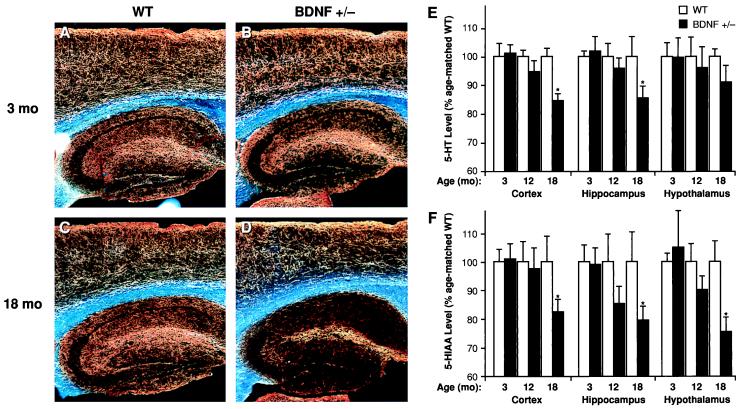
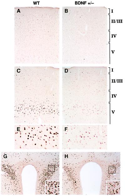
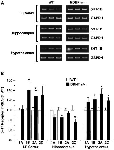
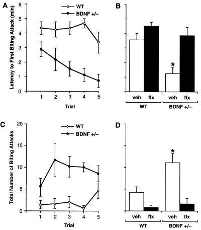
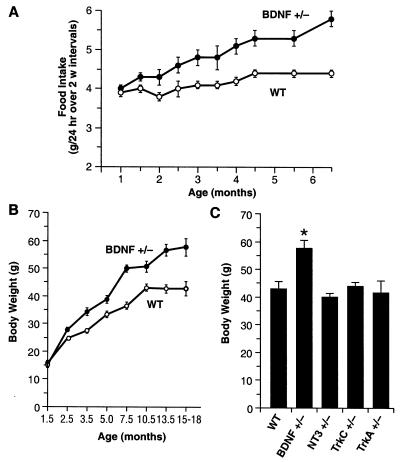
Brain-derived neurotrophic factor (BDNF) has trophic effects on serotonergic (5-HT) neurons in the central nervous system. However, the role of endogenous BDNF in the development and function of these neurons has not been established in vivo because of the early postnatal lethality of BDNF null mice. In the present study, we use heterozygous BDNF(+/-) mice that have a normal life span and show that these animals develop enhanced intermale aggressiveness and hyperphagia accompanied by significant weight gain in early adulthood; these behavioral abnormalities are known to correlate with 5-HT dysfunction. Forebrain 5-HT levels and fiber density in BDNF(+/-) mice are normal at an early age but undergo premature age-associated decrements. However, young adult BDNF(+/-) mice show a blunted c-fos induction by the specific serotonin releaser-uptake inhibitor dexfenfluramine and alterations in the expression of several 5-HT receptors in the cortex, hippocampus, and hypothalamus. The heightened aggressiveness can be ameliorated by the selective serotonin reuptake inhibitor fluoxetine. Our results indicate that endogenous BDNF is critical for the normal development and function of central 5-HT neurons and for the elaboration of behaviors that depend on these nerve cells. Therefore, BDNF(+/-) mice may provide a useful model to study human psychiatric disorders attributed to dysfunction of serotonergic neurons.


Figures

Figure 1
Figure 1
Serotonergic innervation and neurochemical levels of 5-HT and 5-HIAA in the forebrain of BDNF+/− or WT mice. Darkfield photomicrographs (AD) of 5-HT-immunoreactive axons (sagittal sections) in WT (A and C) and BDNF+/− mice (B and D) at 3 months (A and B) or 18 months (C and D) of age. There is a normal 5-HT axon density in neocortex and hippocampus of younger BDNF+/− mice but a loss of axons in 18-month-old BDNF+/− mice relative to WT mice of the same age; we also observed a loss of 5-HT axon density in some of the 12-month-old BDNF+/− mice relative to age-matched WT controls (not shown). The total tissue contents of 5-HT (E) and its metabolite 5-HIAA (F) in the neocortex, hippocampus, and hypothalamus of BDNF+/− and WT littermates at 3, 12, or 18 months of age were measured by HPLC (n = 6–9/group). Data is expressed as a percentage of the mean value found in age-matched WT controls. Repeated measures ANOVA revealed significant reductions in the levels of 5-HT (F1,16 = 7.42, P < 0.05) and 5-HIAA (F1,16 = 8.04, P < 0.05) in 18-month-old BDNF+/− mice relative to WT littermates but not in the younger mutant mice (P > 0.05) in these brain areas. *, P < 0.05.
Figure 2
Figure 2
Induction of c-Fos immunoreactivity 2 h after dFen administration (3 or 10 mg/kg, i.p.) in 3- to 6-month-old WT and BDNF+/− mice (n = 4–5/group). In LF cortex (AF), c-Fos immunoreactivity was low or absent after a vehicle injection in WT (A) or BDNF+/− (B) mice. dFen (10 mg/kg) elicited a robust induction of c-Fos immunoreactivity in the upper part of layer V in LF cortex of WT mice (C; higher magnification shown in E), which is in precise register with a dense band of 5-HT2A/2C receptors and a dense plexus of 5-HT axon terminals found in this zone (43). This induction was blunted in BDNF+/− mice, as indicated by the lower number and staining intensity of c-Fos immunoreactive neurons (D; higher magnification in F). The lower dose of dFen (3 mg/kg) elicited a weak-to-moderate c-Fos induction in LF cortex of WT mice whereas no response was observed in BDNF+/− mice at this dose (not shown). dFen-induced activation of c-Fos was also attenuated in the paraventricular nucleus of the hypothalamus in BDNF+/− mice (H) relative to WT controls (G), a brain area suggested to mediate the anorectic effects of dFen (39).
Figure 3
Figure 3
Reverse transcription–PCR analysis of 5-HT1A, 5-HT1B, 5-HT2A, and 5-HT2C receptor mRNA levels in lateral frontal cortex (LF Cortex), hippocampus, and hypothalamus of WT and BDNF+/− mice. (A) Example of ethidium bromide-stained agarose gels demonstrating 5-HT1B and corresponding glyceraldehyde-3-phosphate dehydrogenase (GAPDH) amplication products (≈330 and 450 bp, respectively) in the indicated brain areas of three WT and three BDNF+/− mice. (B) 5-HT receptor mRNA levels were measured in WT and BDNF+/− mice (6–9 months of age) by reverse transcription–PCR (see Materials and Methods; n = 6–7/group). There were significant (ANOVA, P < 0.05) up-regulations of 5HT1B and 5HT2A receptors in LF cortex and hypothalamus, an up-regulation of the 5HT2C receptor in hypothalamus, and a down-regulation of the 5HT2C receptor in the hippocampus of BDNF+/− mice relative to WT controls; a marginal (P = 0.05–0.1) up-regulation of the 5HT2C receptor was found in LF cortex, and marginal down-regulations of the 5HT1A and 5HT1B receptors were found in the hippocampus of BDNF+/− mice. *, P < 0.05.
Figure 4
Figure 4
Offensive intermale aggression in BDNF+/− and WT littermates as assessed in a resident-intruder assay (n = 8–12/group; 2.5–4.5 months of age). (A) Latency to first biting attack. (C) Number of biting attacks measured during five consecutive trials. Repeated measures ANOVA revealed significant differences between the BDNF+/− and WT groups for both biting attack latency (F1,14 = 13.67, P < 0.005) and number of attacks (F1,14 = 9.98, P < 0.01), evident as early as the first trial. (B and D) Chronic Flx treatment in BDNF+/− mice (BDNF+/− flx) normalized both the attack latency (B) and number of attacks (D) to levels found in WT mice administered either vehicle (WT veh) or Flx (WT flx). *, P < 0.05 (ANOVA; different from all other treatment groups whereas no differences were found between the other three groups).
Figure 5
Figure 5
BDNF+/− mice develop hyperphagia and elevated body mass. (A) Daily food intake (g/day) of BDNF+/− and WT mice (n = 12–13/group). Values indicate the mean ± SEM of daily food intake averaged over a 2-week interval. Food intake of BDNF+/− mice was significantly higher than in age-matched WT controls (ANOVA, P < 0.05). (B) Growth curves for BDNF+/− and WT mice (n = 110–115/group) indicate that BDNF+/− mice have a significantly higher body weight than WT littermates, beginning at 10 weeks of age (ANOVA, P < 0.001). (C) Body weight of 15-month-old WT, BDNF+/−, and mice heterozygous for other members of the neurotrophin (NT-3+/−) or neurotrophin receptor family (trkA+/− and trkC+/−). *, P < 0.05 compared with WT.

References

    1. Eaton M J, Staley J K, Globus M Y, Whittemore S R. Dev Biol. 1995;170:169–182. - PubMed
    1. Mamounas L A, Blue M E, Siuciak J A, Altar C A. J Neurosci. 1995;15:7929–7939. - PMC - PubMed
    1. Siuciak J A, Boylan C, Fritsche M, Altar C A, Lindsay R M. Brain Res. 1996;710:11–20. - PubMed
    1. Baumgarten H G, Grozdanovic Z. Pharmacopsychiatry. 1995;28, Suppl. 2:73–79. - PubMed
    1. Hen R. Neuron. 1996;16:17–21. - PubMed

Publication types

LinkOut - more resources