. 1999 Dec 21;96(26):15268-73.
doi: 10.1073/pnas.96.26.15268.

Abnormal neurotransmission in mice lacking synaptic vesicle protein 2A (SV2A)

Affiliations

Abnormal neurotransmission in mice lacking synaptic vesicle protein 2A (SV2A)

K M Crowder et al. Proc Natl Acad Sci U S A. .

Abstract

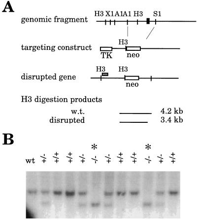
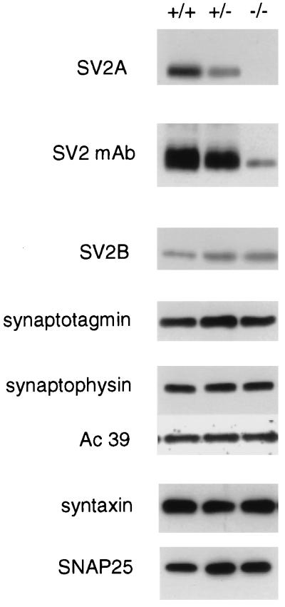
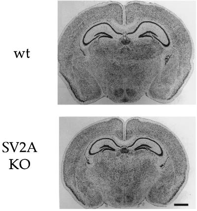
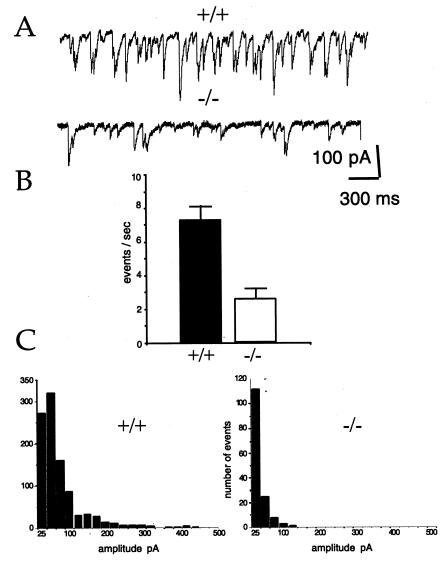
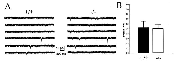
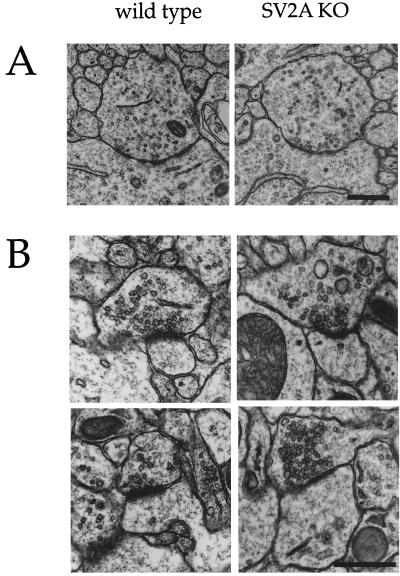
Synaptic vesicle protein 2 (SV2) is a membrane glycoprotein common to all synaptic and endocrine vesicles. Unlike many proteins involved in synaptic exocytosis, SV2 has no homolog in yeast, indicating that it performs a function unique to secretion in higher eukaryotes. Although the structure and protein interactions of SV2 suggest multiple possible functions, its role in synaptic events remains unknown. To explore the function of SV2 in an in vivo context, we generated mice that do not express the primary SV2 isoform, SV2A, by using targeted gene disruption. Animals homozygous for the SV2A gene disruption appear normal at birth. However, they fail to grow, experience severe seizures, and die within 3 weeks, suggesting multiple neural and endocrine deficits. Electrophysiological studies of spontaneous inhibitory neurotransmission in the CA3 region of the hippocampus revealed that loss of SV2A leads to a reduction in action potential-dependent gamma-aminobutyric acid (GABA)ergic neurotransmission. In contrast, action potential-independent neurotransmission was normal. Analyses of synapse ultrastructure suggest that altered neurotransmission is not caused by changes in synapse density or morphology. These findings demonstrate that SV2A is an essential protein and implicate it in the control of exocytosis.


Figures

Figure 1
Figure 1
Targeted disruption of the SV2A gene. (A) Strategy for generating SV2A-minus mice. A portion of the SV2A gene was isolated by screening a mouse 129SV genomic DNA library with a probe corresponding to the 5′-end of the rat SV2A cDNA. The exon containing the translation start site is depicted as a black box. A targeting construct was generated in which this exon and surrounding DNA were replaced with a gene encoding neomycin resistance. To allow for negative selection, a gene encoding thymidine kinase was placed at the end of the short arm of the construct. Homologous recombination of the construct resulted in a disrupted gene that produced a shorter fragment when digested with the restriction enzyme HindIII. Cells and animals were genotyped by Southern analysis of HindIII-digested genomic DNA probed with the DNA fragment depicted as a gray box above the schematic of the disrupted gene.H3, HindIII; X1, XbaI; A1, ApaI; S1, SpeI; neo, gene encoding neomycin resistance; TK, gene encoding thymidine kinase; w.t., wild type. (B) Southern analysis demonstrating disruption of the SV2A gene. Shown is a Southern analysis of genomic DNA isolated from two litters produced by mice heterozygous (+/−) for the SV2A gene disruption. DNA containing the disruption produces a smaller HindIII-digestion fragment. Genotypes are indicated at the tops of the lanes. Homozygous mutants are marked with an asterisk. wt, wild type.
Figure 2
Figure 2
Disruption of the SV2A gene results in the absence of SV2A protein but does not significantly alter the expression of other synaptic proteins. Shown are immunoblot analyses of brain protein isolated from wild-type (+/+) mice and littermates heterozygous (+/−) and homozygous (−/−) for the SV2A mutation. SV2A expression was detected by using an isoform-specific polyclonal antibody. Mice heterozygous for the mutation express significantly less SV2A than wild-type littermates, and homozygous mutants express nondetectable levels. Western analysis using the anti-SV2 monoclonal antibody, which recognizes an epitope present in all known SV2 isoforms, revealed that SV2A knockouts (−/−) express very little total SV2 protein. This suggests that SV2A is the primary SV2 isoform in mouse brain. This decrease is seen even though the expression of SV2B is increased in these animals. Expression levels were normal for the synaptic proteins synaptotagmin I, synaptophysin, and the 39-kDa accessory subunit of the H+/ATPase (Ac 39), and for the t-SNAREs syntaxin and SNAP-25.
Figure 3
Figure 3
Gross brain morphology is normal in SV2A-knockout mice. Shown are Nissl-stained coronal sections of brain from P14 wild-type and SV2A-knockout mice. The brains of SV2A-knockout mice are smaller, in proportion with their reduced body size. However, major brain structures, including the cerebral cortex, hippocampus, and thalamus, appear normal. Cerebellar morphology was also normal (data not shown). wt, wild type; KO, knockout.
Figure 4
Figure 4
GABAergic neurotransmission is reduced in the hippocampus of SV2A-knockout animals. sIPSCs were measured in hippocampal CA3 pyramidal neurons. Recordings were made in the presence of the glutamate receptor blockers CNQX and APV. (A) Sample traces from wild-type (+/+) and SV2A-knockout (−/−) cells. Shown are 4-sec traces of sIPSCs recorded from representative cells. The downward deflection of the events is the result of outward chloride flux in the cells voltage-clamped at −70 mV. Currents were filtered at 1 kHz and digitized at 5 kHz. There was no digital filtering of the recordings to decrease noise in the traces shown. (B) Averaged sIPSC frequencies. Shown are histograms representing average sIPSC frequencies. Each was calculated from 30 2-min epochs. Wild-type values were obtained from six cells from six (+/+) animals. Knockout values were obtained from seven cells from five (−/−) animals. Error bars indicate SEM. Rates were significantly different as determined with the Student's t test (P ≪ 0.01). (C) Frequency histograms of sIPSC amplitude. A frequency analysis of event amplitudes binned in 25-pA increments is shown. Data were derived from recordings of six cells from six (+/+) animals and seven cells from four (−/−) animals. For each cell, a single 2-min epoch of representative frequency was selected. All events detected by the detection program were verified by visual inspection. Note that there were many fewer events in (−/−) cells. The average amplitude in (−/−) cells was roughly half that of events in (+/+) cells, 24.85 pA ± 7.8 vs. 51.2 pA ± 6.8 for the knockouts and wild type, respectively (n = 4 each).
Figure 5
Figure 5
Action potential-independent neurotransmission is not affected in SV2A-knockout animals. (A) Sample traces from wild-type (+/+) and SV2A-knockout (−/−) cells. Shown are representative 23-sec sweeps of mIPSCs from whole-cell voltage clamp recordings of hippocampal CA3 pyramidal cells held at −70 mV. Recordings were made in the presence of the glutamate receptor blockers APV and CNQX and the sodium channel blocker tetrototoxin. (B) mIPSC frequencies were similar in cells from wild type and SV2A knockouts. Histograms illustrating the average frequency of mIPSCs recorded from nine (+/+) cells (six animals) and nine (−/−) cells (six animals). Error bars indicate SEM. Rates were not significantly different as determined with the Student's t test (P = 0.79).
Figure 6
Figure 6
Synapse morphology is normal in SV2A-knockout mice. (A) Shown are representative electron micrographs of symmetric (GABAergic) synapses in the cell-body region of CA3 hippocampal neurons. These synapses are presumed to be GABAergic based on their location, symmetry of active zones, and irregularly shaped vesicles. Qualitative inspection of these synapses suggested no difference in density or morphology between wild type and SV2A knockouts. (B) Shown are representative electron micrographs of asymmetric synapses onto proximal dendrites of CA3 hippocampal neurons. Synapses in this region are primarily from dentate granule cells and are largely asymmetric, presumed glutamatergic synapses. Similar morphology was observed in wild type and SV2A knockouts. Quantitative analyses of these synapses are presented in Table 1.

References

    1. Bajjalieh S M. Curr Opin Neurobiol. 1999;9:321–328. - PubMed
    1. Buckley K, Kelly R B. J Cell Biol. 1985;100:1284–1294. - PMC - PubMed
    1. Bajjalieh S M, Peterson K, Shinghal R, Scheller R H. Science. 1992;257:1271–1273. - PubMed
    1. Feany M B, Lee S, Edwards R H, Buckley K M. Cell. 1992;70:861–867. - PubMed
    1. Bajjalieh S M, Peterson K, Linial M, Scheller R H. Proc Natl Acad Sci USA. 1993;90:2150–2154. - PMC - PubMed

Publication types

MeSH terms