Comparative Study
. 1999 Dec 21;96(26):15348-53.
doi: 10.1073/pnas.96.26.15348.

A bZIP factor, TRAB1, interacts with VP1 and mediates abscisic acid-induced transcription

Affiliations
Comparative Study

A bZIP factor, TRAB1, interacts with VP1 and mediates abscisic acid-induced transcription

T Hobo et al. Proc Natl Acad Sci U S A. .

Abstract

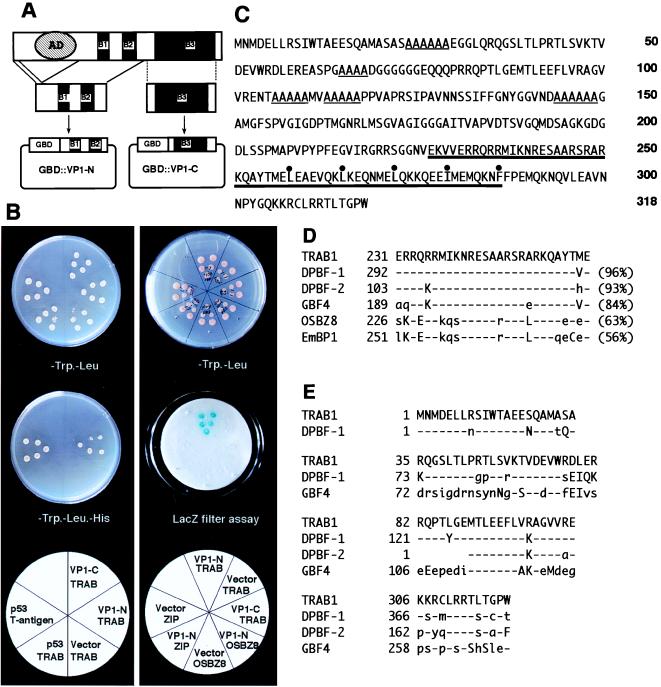
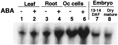
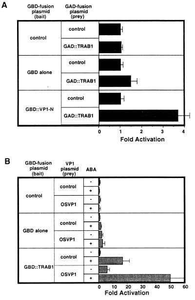
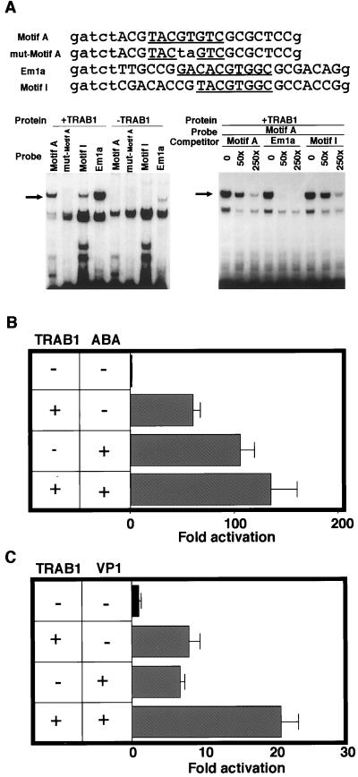
The transcription factor VP1 regulates maturation and dormancy in plant seeds by activating genes responsive to the stress hormone abscisic acid (ABA). Although activation involves ABA-responsive elements (ABREs), VP1 itself does not specifically bind ABREs. Instead, we have identified and cloned a basic region leucine zipper (bZIP) factor, TRAB1, that interacts with both VP1 and ABREs. Transcription from a chimeric promoter with GAL4-binding sites was ABA-inducible if cells expressed a GAL4 DNA-binding domain::TRAB1 fusion protein. Results indicate that TRAB1 is a true trans-acting factor involved in ABA-regulated transcription and reveal a molecular mechanism for the VP1-dependent, ABA-inducible transcription that controls maturation and dormancy in plant embryos.


Figures

Figure 1
Figure 1
Isolation and characterization of TRAB1 cDNA. (A) Schematic illustration of the bait construct (pGBD∷VP1-N) used for yeast two-hybrid screening. pGBD∷VP1-C was used as a control bait. The conserved basic regions (B1, B2, and B3) and the acidic transcriptional activation domain of OSVP1 are indicated by solid boxes and a hatched oval, respectively. (B) Yeast two-hybrid assays showing that TRAB1 specifically interacts with the VP1-N fragment. (Left) HIS3 assays. Five independent yeast transformants harboring various combinations of a bait (GBD fusion) and prey (GAD fusion) plasmids were grown on an SD medium plate containing 30 mM 3-aminotriazole, lacking Trp and Leu (Top) or lacking Trp, Leu, and His (Middle). At the bottom, the combination of bait (top line) and prey (bottom line) fragments fused to GBD and GAD, respectively, is indicated in each sector. The combination of the T antigen prey and p53 bait was used as a control for positive interaction. “Vector” indicates a negative control bait of GBD alone (pGBT9). Note that only transformants with the combination of pGBD∷VP1-N and pGAD∷TRAB1 or the positive controls grew on the minus His medium. (Right) LacZ assays. Yeast transformants harboring the indicated combinations of plasmids were grown on an SD medium plate lacking Trp and Leu (Top), and replicate filters were used for LacZ filter assays with X-Gal (Middle). OSBZ8 (12) and ZIP are negative control preys. The cDNA for ZIP was obtained by a yeast one-hybrid screen of the same cDNA library by using the 55-bp ABRC of Osem promoter as a target site and found to encode a protein with a bZIP structure very similar to that of TRAB1 in-frame with GAD. Note that positive signals were obtained only with the combination of GBD∷VP1-N and GAD∷TRAB1. (C) Amino acid sequence of TRAB1 deduced from the nucleotide sequence of the cDNA. The bZIP region and short stretches of Ala residues are indicated by thick and thin underlines, respectively. Hydrophobic amino acid residues that constitute a leucine zipper structure are marked with dots. (D) Comparison between the sequence of the basic region of TRAB1 and those of DPBF-1, DPBF-2, GBF4, OSBZ8, and EmBP1. Percentages of identical amino acids to those of TRAB1 are shown in parentheses. Amino acids identical, similar, and dissimilar to those of TRAB1 are indicated by dashes and uppercase and lowercase letters, respectively. (E) Amino acid sequence blocks conserved between TRAB1, DPBF-1, DPBF-2, and GBF4. Amino acids identical, similar, and dissimilar to those of TRAB1 are indicated by dashes and uppercase and lowercase letters, respectively.
Figure 2
Figure 2
Northern hybridization analysis of TRAB1. Thirty micrograms of RNA was subjected to Northern hybridization analysis by using a TRAB1 cDNA probe. RNA was from leaves (lanes 1 and 2) and roots (lanes 3 and 4) of young rice plants (14 days old) treated with 10−4 M ABA for 6 h (lanes 2 and 4) or receiving no ABA (lanes 1 and 3), suspension-cultured cells (Oc cells) treated with 5 × 10⁻⁵ M ABA for 6 h (lane 6) or receiving no ABA (lane 5), and rice embryos at 13–14 days after flowering (lane 7) or at dry mature stage (lane 8). The entire TRAB1 cDNA was used for the probe.
Figure 3
Figure 3
Interaction between TRAB1 and VP1 in rice protoplast cells. (A) Two-hybrid interactions in the plant cell. Ten micrograms each of bait plasmid [p35S-ShΔ-stop (control), p35S-ShΔ-GBD (GBD alone), or p35S-ShΔ-GBD∷VP1-N (GBD∷VP1-N)], prey plasmid [p35S-ShΔ-stop (control) or p35S-ShΔ-GAD∷TRAB1 (GAD∷TRAB1)], and the UAS-TATA-GUS reporter plasmid were cotransfected into protoplasts of rice suspension-cultured cells by electroporation. The electroporated cells were cultured for 40 h. The control plasmid (p35S-ShΔ-stop) consisted of CaMV 35S promoter, Sh1 first intron with an internal deletion, and NOS terminator, but no coding region. Each transfection also included 5 μg of a ubiquitin promoter∷luciferase plasmid as an internal standard. GUS activities were normalized by luciferase activity. The effects of GAD∷TRAB1 (prey) fusion on the expression of the reporter gene are expressed by fold activation of normalized GUS activities relative to that obtained with a combination of indicated bait and control plasmids. Each value is the mean of four replicate electroporations with standard error. (B) Interaction between VP1 and GBD∷TRAB1 and the capacity of GBD∷TRAB1 fusion to confer ABA responsiveness to a promoter carrying GAL4-binding sites. Ten micrograms each of bait (GBD fusion) plasmid [p35S-ShΔ-stop (control), p35S-ShΔ-GBD (GBD alone), or p35S-ShΔ-GBD∷TRAB1 (GBD∷TRAB1], prey plasmid [p35-S-ShΔ-stop (control) or p35-S-Sh-OSVP1 (OSVP1)], and the UAS-TATA-GUS reporter plasmid were cotransfected into protoplasts of rice suspension-cultured cells by electroporation, and the cells were cultured for 40 h in the absence (−) or presence (+) of 5 × 10⁻⁵ M ABA. The effects of OSVP1 and ABA on the expression of the reporter gene are expressed by fold activation of normalized GUS activities relative to that obtained with a combination of indicated GBD (bait) fusion and control plasmid in the absence of ABA. Other details are as described for A.
Figure 4
Figure 4
Interaction of TRAB1 with ABREs and activation of the ABRC of Osem promoter consisting of ABRE and CE3. (A) Electrophoretic mobility-shift assays of TRAB1 synthesized in vitro. Nucleotide sequences of ABREs (underlined) and its mutant used for probes of electrophoretic mobility-shift assays are listed on the top. The synthetic double-strand DNA fragments carried 4-bp 5′ overhangs at each end for fill-in labeling. Each different probe without specific competitors (Left) or motif A probe with an unlabeled competitor as indicated (Right) was incubated with TRAB1 protein (+TRAB1) synthesized in a rabbit reticulocyte cell-free system. “−TRAB1” indicates the control binding reactions with the reticulocyte lysate after protein synthesis reaction without added RNA templates. “×” indicates fold-molar excess. DNA–protein complexes were separated from free probes by PAGE. Arrows indicate the position of the bands of DNA–protein complexes specific to TRAB1 protein. Other bands derive from the protein present in the reticulocyte lysate. (B) Activation of the 55-bp ABRC of Osem by TRAB1. Ten micrograms of the ABRC-TATA-GUS reporter plasmid was cotransfected with the same amount of TRAB1 expression (p35S-ShΔ-TRAB1) or control (p35S-ShΔ-stop) plasmids, as indicated, into rice cell protoplasts by electroporation, and protoplasts were cultured in the absence (−) or presence (+) of 5 × 10⁻⁵ M ABA. The effects of TRAB1 and ABA are indicated by fold activation of normalized GUS activities relative to that obtained with the control plasmid in the absence of ABA. Other details are as described for Fig. 3. (C) The effect of VP1 overexpression on the activation of the 55-bp ABRC by TRAB1. Cotransfection experiments were conducted as in B except for the following: the amount of p35S-ShΔ-TRAB1 or its control plasmid was reduced to 5 μg; 30 μg of VP1 expression plasmid (p35S-Sh-OSVP1) or its corresponding control plasmid was included; and protoplasts were cultured only in the absence of ABA.

References

    1. Giraudat J, Parcy F, Bertauche N, Gosti F, Leung J, Morris P C, Bouvier-Durand M, Vartanian N. Plant Mol Biol. 1994;26:1557–1577. - PubMed
    1. Finkelstein R R, Tenbarge K M, Shumway J E, Crouch M L. Plant Physiol. 1985;78:630–636. - PMC - PubMed
    1. McCarty D R. Annu Rev Plant Physiol Plant Mol Biol. 1995;46:71–93.
    1. Busk P K, Pages M. Plant Mol Biol. 1998;37:425–435. - PubMed
    1. Hattori T, Terada T, Hamasuna S. Plant J. 1995;7:913–925. - PubMed

Publication types

MeSH terms

Associated data

LinkOut - more resources