. 2000 Jan;182(1):57-66.
doi: 10.1128/JB.182.1.57-66.2000.

The VirR response regulator from Clostridium perfringens binds independently to two imperfect direct repeats located upstream of the pfoA promoter

Affiliations

The VirR response regulator from Clostridium perfringens binds independently to two imperfect direct repeats located upstream of the pfoA promoter

J K Cheung et al. J Bacteriol. 2000 Jan.

Erratum in

  • J Bacteriol 2000 May;182(10):2992

Abstract

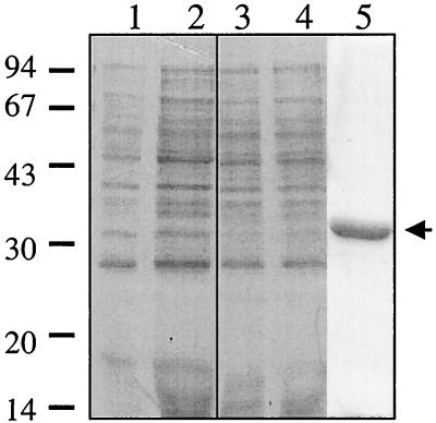
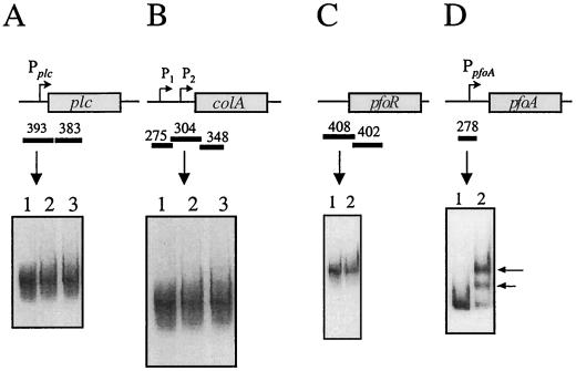
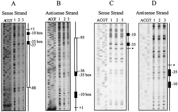
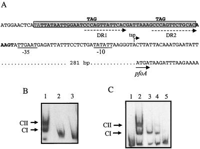
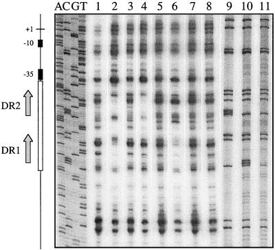
Regulation of toxin production in the gram-positive anaerobe Clostridium perfringens occurs at the level of transcription and involves a two-component signal transduction system. The sensor histidine kinase is encoded by the virS gene, while its cognate response regulator is encoded by the virR gene. We have constructed a VirR expression plasmid in Escherichia coli and purified the resultant His-tagged VirR protein. Gel mobility shift assays demonstrated that VirR binds to the region upstream of the pfoA gene, which encodes perfringolysin O, but not to regions located upstream of the VirR-regulated plc, colA, and pfoR genes, which encode alpha-toxin, collagenase, and a putative pfoA regulator, respectively. The VirR binding site was shown by DNase I footprinting to be a 52-bp core sequence situated immediately upstream of the pfoA promoter. When this region was deleted, VirR was no longer able to bind to the pfoA promoter. The binding site was further localized to two imperfect direct repeats (CCCAGTTNTNCAC) by site-directed mutagenesis. Binding and protection analysis of these mutants indicated that VirR had the ability to bind independently to the two repeated sequences. Based on these observations it is postulated that the VirR positively regulates the synthesis of perfringolysin O by binding directly to a region located immediately upstream of the pfoA promoter and activating transcription.


Figures

FIG. 1
FIG. 1
Purification of VirR. Low-molecular-weight standards (Pharmacia Biotech), in kilodaltons, are shown adjacent to the gel. Lanes 1 and 3, uninduced whole-cell extracts from cells harboring the vector pRSET A or pJIR1342, respectively; lanes 2 and 4, postinduction (4 h) whole-cell extracts from cells carrying the vector or pJIR1342, respectively; lane 5, His-VirR purified from cells induced for 30 to 60 min with IPTG (arrow).
FIG. 2
FIG. 2
Gel mobility shift analysis of target gene regions. Shown are the results of gel mobility shift assays using plc (A), colA (B), pfoR (C), and pfoA (D) gene regions. These regions are shown in the schematic above each gel shift result, with the rectangular boxes representing the respective genes. The direction of transcription is shown by arrows from each promoter (P). The DNA fragments used in the assays are shown by the solid bars, and their respective sizes (in base pairs) are shown above the bars. (A and B) Lane 1, no-VirR control; lanes 2 and 3, DIG-labelled target DNA incubated with 1 and 2 μg of VirR, respectively. (C) Lane 1, no-VirR control; lane 2, target DNA incubated with 1 μg of VirR. (D) Lanes 1 and 2, 278-bp fragment incubated with no protein or 1 μg of VirR, respectively. Shifted bands are indicated by the arrows.
FIG. 3
FIG. 3
Gel shift analysis of the pfoA gene region. The pfoA-derived DNA fragments used in the gel mobility shift assays (bars) and their respective sizes (in base pairs) are shown. The direct repeats in the 114-bp fragment, the promoter in the 183-bp fragment, and the inverted repeats in the 211-bp fragment, are shown as directly repeated arrows, a bent arrow (PpfoA), and inverted arrows, respectively. (A and B) Lane 1, no-VirR control; lanes 2 and 3, DIG-labelled DNA incubated with 1 and 2 μg of VirR, respectively. The VirR-DNA complexes CI and CII are indicated. (C) Lane 1, no-VirR control; lane 2, DIG-labelled fragment incubated with 1 μg of VirR. (D) Lane 1, no-VirR control; lanes 2 to 5, target DNA incubated with 1 μg of VirR. Reaction mixtures in lanes 3 to 5 also contained 15 pmol of the following unlabelled fragments: lane 3, 302-bp fragment (specific competitor); lane 4, 183-bp of unlabelled upstream pfoA fragment; lane 5, 408-bp upstream pfoR fragment (nonspecific competitor).
FIG. 4
FIG. 4
Specificity and VirR dependence of DNA binding. (A) Competitive binding gel shift assay. The DIG-labelled 183-bp pfoA fragment was incubated with 1 μg of VirR and various amounts of unlabelled 183-bp DNA (specific competitor). Lane 1, no-VirR control; lanes 2 to 6, target DNA that was incubated with 1 μg of VirR. These incubation mixtures also contained 0, 3.0, 7.5, 15, and 30 pmol of specific competitor DNA, respectively. Lane 7, DIG-labelled DNA incubated with 1 μg of VirR and 30 pmol of nonspecific competitor (408-bp upstream pfoR fragment). (B) Concentration dependence of VirR binding. The [α-32P]dATP-labelled 183-bp pfoA fragment was incubated with various amounts of VirR and examined by gel shift analysis as described above except that the data were obtained and quantitated in a phosphorimager. The amount of labelled DNA in each band was calculated and plotted as shown. The amounts of free DNA (F), CI complex, CII complex, and total complex (CI+CII) are shown.
FIG. 5
FIG. 5
DNase I footprinting analysis. Results of footprinting reactions where either the sense or antisense DNA strands were labelled with [γ-32P]ATP are shown. Sequencing reactions with the same oligonucleotide primers used to generate the PCR products are shown next to the footprinting reactions. The −10 and −35 boxes are represented by the black rectangles. (A and B) Identification of the VirR binding site. Regions protected from DNase I digestion are represented by the open rectangles. The transcription start point is shown as +1, and the positions of the regions of protection relative to +1 are as indicated. (C and D) Analysis of the VirR binding site deletion derivative. The site of deletion is indicated by the asterisk. In all panels, lane 1 contains no VirR and lanes 2 and 3 contain 1 and 2 μg of VirR, respectively. All footprinting reaction mixtures contained 25,000 cpm of labelled DNA.
FIG. 6
FIG. 6
Sequence and mutations of the VirR binding site. (A) Sequence of the core binding region site (boldface). The imperfect direct repeats within this region are indicated by the dashed arrows. The nucleotide residues that were changed by site-directed mutagenesis are shown above the original nucleotide sequence. The −35 and −10 boxes of the pfoA promoter are underlined, the transcriptional start point (tsp) is indicated by the bent arrow, and the start of the pfoA gene is indicated by the solid arrow. The 49-bp region deleted by SOE PCR is shown as the gray rectangle. (B) Gel mobility shift assays carried out on the deletion derivatives. Lane 1, wild-type 183-bp fragment incubated with 1 μg of VirR; lanes 2 and 3, 134-bp deletion fragment incubated with either no VirR or 1 μg of VirR, respectively. (C) Gel mobility shift assay with direct repeat mutation derivatives. The imperfect direct repeats were altered by site-directed mutagenesis. Lanes 1 and 2, DNA fragment with intact direct repeats; lanes 3 to 5, DNA fragments with mutations in DR1, DR2, or DR1 and DR2, respectively. All binding reaction mixtures contained 1 μg of VirR, with the exception of the no-protein control in lane 1.
FIG. 7
FIG. 7
DNase I footprinting analysis of direct-repeat mutants. The labelled plasmid templates used in this experiment were the wild-type plasmid pJIR1546 (lanes 1 and 2) and the mutated derivatives pJIR1804 (DR1 mutant; lanes 3 and 4), pJIR1803 (DR2 mutant; lanes 5 and 6), and pJIR1821 (DR1-DR2 double mutant; lanes 7 and 8). Lanes 1, 3, 5, and 7, control reaction mixtures that were not preincubated with VirR. Lanes 2, 4, 6, and 8, test reaction mixtures that were incubated with 2 μg of VirR prior to partial digestion by DNase I and electrophoresis. The first four lanes (ACGT) show the sequencing reaction products from the wild-type pJIR1546 template. Lanes 9 to 11, C track sequencing reaction products from pJIR1804, pJIR1803, and pJIR1821, respectively. The positions of the DR1 and DR2 repeats are shown by the arrows, and the region of protection is represented by the open rectangle. The −10 and −35 boxes are depicted as black rectangles, and the transcription start point is shown as +1.
FIG. 8
FIG. 8
Proposed model of the VirS/VirR two-component signal transduction system. The genes postulated to be controlled by the VirS/VirR network are shown as light gray boxes. The dark gray rectangles represent the VirS sensor histidine kinase, while the VirR response regulator is depicted by the dark gray ovals. The presence of other activator(s) is symbolized by the black oval. The unknown protease and sialidase genes are represented by prt and nan, respectively.

References

    1. Albright L M, Huala E, Ausubel F M. Prokaryotic signal transduction mediated by sensor and regulator protein pairs. Annu Rev Genet. 1989;23:311–336. - PubMed
    1. Awad M M, Bryant A E, Stevens D L, Rood J I. Virulence studies on chromosomal α-toxin and θ-toxin mutants constructed by allelic exchange provide genetic evidence for the essential role of α-toxin in Clostridium perfringens-mediated gas gangrene. Mol Microbiol. 1995;15:191–202. - PubMed
    1. Ba-Thein W, Lyristis M, Ohtani K, Nisbet I T, Hayashi H, Rood J I, Shimizu T. The virR/virS locus regulates the transcription of genes encoding extracellular toxin production in Clostridium perfringens. J Bacteriol. 1996;178:2514–2520. - PMC - PubMed
    1. Bellsolell L, Prieto J, Serrano L, Coll M. Magnesium binding to the bacterial chemotaxis protein CheY results in large conformational changes involving its functional surface. J Mol Biol. 1994;238:489–495. - PubMed
    1. Boucher P E, Menozzi F D, Locht C. The modular architecture of bacterial response regulators. Insights into the activation mechanism of the BvgA transactivator of Bordetella pertussis. J Mol Biol. 1994;241:363–377. - PubMed

Publication types

MeSH terms

LinkOut - more resources