. 2000 Jan;182(1):155-63.
doi: 10.1128/JB.182.1.155-163.2000.

Genetics of L-sorbose transport and metabolism in Lactobacillus casei

Affiliations

Genetics of L-sorbose transport and metabolism in Lactobacillus casei

M J Yebra et al. J Bacteriol. 2000 Jan.

Abstract

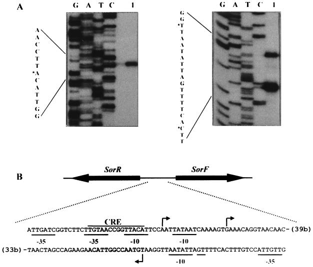
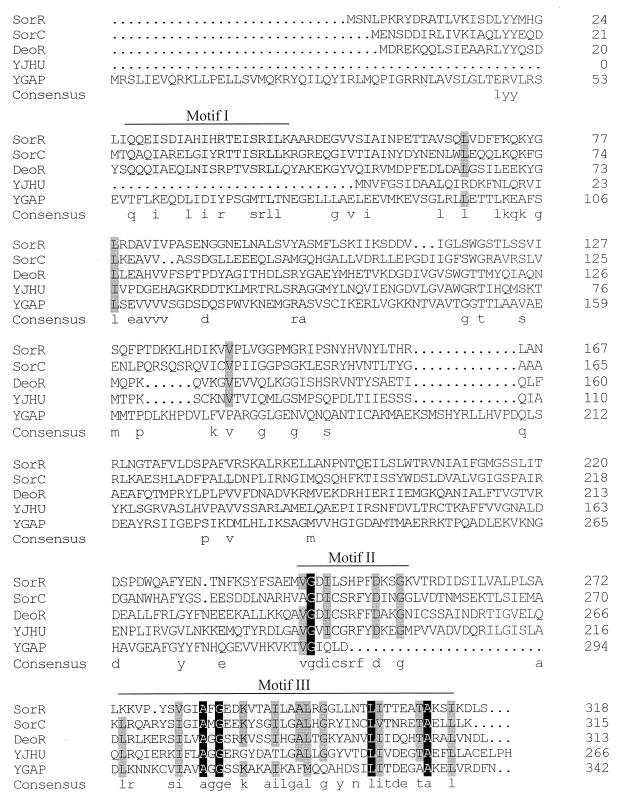
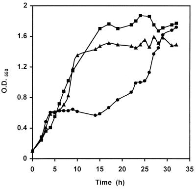
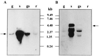
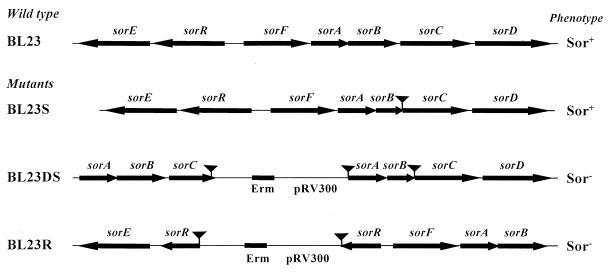
Genes encoding L-sorbose metabolism of Lactobacillus casei ATCC 393 have been identified on a 6.8-kb chromosomal DNA fragment. Sequence analysis revealed seven complete genes and a partial open reading frame transcribed as two units. The deduced amino acid sequences of the first transcriptional unit (sorRE) showed high similarity to the transcriptional regulator and the L-sorbose-1-phosphate reductase of the sorbose (sor) operon from Klebsiella pneumoniae. The other genes are transcribed as one unit (sorFABCDG) in opposite direction to sorRE. The deduced peptide sequence of sorF showed homology with the D-sorbitol-6-phosphate dehydrogenase encoded in the sor operon from K. pneumoniae and sorABCD to components of the mannose phosphotransferase system (PTS) family but especially to domains EIIA, EIIB, EIIC and EIID of the phosphoenolpyruvate-dependent L-sorbose PTS from K. pneumoniae. Finally, the deduced amino acid sequence of a truncated gene (sorG) located downstream of sorD presented high similarity with ketose-1,6-bisphosphate aldolases. Results of studies on enzyme activities and transcriptional analysis revealed that the two gene clusters, sorRE and sorFABCDG, are induced by L-sorbose and subject to catabolite repression by D-glucose. Data indicating that the catabolite repression is mediated by components of the PTS elements and by CcpA, are presented. Results of sugar uptake assays in L. casei wild-type and sorBC mutant strains indicated that L-sorbose is taken up by L-sorbose-specific enzyme II and that L. casei contains an inducible D-fructose-specific PTS. Results of growth analysis of those strains and a man sorBC double mutant suggested that L-sorbose is probably also transported by the D-mannose PTS. We also present evidence, from studies on a sorR mutant, suggesting that the sorR gene encodes a positive regulator of the two sor operons. Sequence alignment of SorR, SorC (K. pneumoniae), and DeoR (Bacillus subtilis) revealed that they might constitute a new group of transcriptional regulators.


Figures

FIG. 1
FIG. 1
(A) Schematic representation of the transport and metabolism of l-sorbose. (B) Simplified restriction map and genetic organization of the sorbose operons from L. casei.
FIG. 2
FIG. 2
(A) Primer extension signals for sorRE (left) and sorFABCDG (right) promoters. Lanes G, A, T, and C contain the DNA sequencing reactions; lane 1 contains the products of primer extension. The DNA sequence shown is the complementary strand of the mRNA. Asterisks indicate nucleotides corresponding to the main extension products. (B) Sequence of the sor promoter region. The transcriptional start sites of the sorRE and sorFABCDG promoters are indicated by arrows. The putative promoter sequences, −35 and −10, are underlined. The cre-like sequence is also shown. Numbers in parentheses indicate distances in nucleotides from the sorR and sorF ATG start codons.
FIG. 3
FIG. 3
Multiple amino acid sequence alignment of SorR from L. casei (this work), SorC from K. pneumoniae (accession no. P37078), DeoR from B. subtilis (accession no. P39140), YJHU from E. coli (accession no. P39356), and YGAP from B. megaterium (accession no. P35168). The residue number of each protein is indicated at the right. The consensus sequence (at least three residues conserved) is shown in lowercase letters. Residues conserved in all proteins are shown against a dark background. Residues that are similar in four sequences appear against a shaded background. Motif I indicates the position of the putative DNA-binding sites; motifs II and III indicate highly conserved regions.
FIG. 4
FIG. 4
Growth of L. casei on MRS basal medium containing 0.1% glucose plus 0.2% sorbose. ●, wild type (BL23); ▴, ccpA mutant (BL71); ■, man mutant (BL23D).
FIG. 5
FIG. 5
Northern blot analysis of sor mRNA, using an internal DNA fragment of sorE (A) or sorF (B) as the probe. RNA was isolated from L. casei grown with 0.5% glucose (g), 0.5% sorbose (s), 0.5% glucose plus 0.5% sorbose (gs), or 0.5% ribose (r). Arrows indicate transcripts of about 2,200 and 4,400 nucleotides; positions of size standards are marked in the center.
FIG. 6
FIG. 6
Schematic representation of the genetic organization of the sorbose operons in wild-type and mutant strains of L. casei. The strategy for disrupting the sor genes is described in Materials and Methods. Arrows indicate ORFs and their orientations. Deleted DNA fragments are represented by triangular symbols. The erythromycin gene (Erm) and DNA from plasmid pRV300 are also indicated. Phenotypes were tested by streaking single colonies on MRS fermentation medium with 0.5% l-sorbose. + and − indicate fermentation and no fermentation, respectively.
FIG. 7
FIG. 7
(A) d-[14C]fructose uptake rate in L. casei wild-type (BL23) and sorBC mutant strains. Cells were pregrown on MRS fermentation medium with 0.5% fructose (■, wild type; □, sorBC mutant) or 0.5% sorbose (●, wild type; ○, sorBC mutant). (B) Lineweaver-Burk plot of d-[14C]fructose uptake rate without sorbose (●) or with sorbose (■, 10 μM; ▴, 100 μM) by the wild type pregrown on MRS fermentation medium with 0.5% sorbose.

References

    1. Alpert C-A, Siebers U. The lac operon of Lactobacillus casei contains lacT, a gene coding for a protein of the BglG family of transcriptional antiterminators. J Bacteriol. 1997;179:1555–1562. - PMC - PubMed
    1. Bradford M M. A rapid and sensitive method for the quantitation of microgram quantities of protein utilizing the principle of protein-dye binding. Anal Biochem. 1976;72:248–254. - PubMed
    1. Burland V, Plunkett III G, Sofia H J, Daniels D L, Blattner F R. Analysis of the Escherichia coli genome VI: DNA sequence of the region from 92.8 through 100 minutes. Nucleic Acids Res. 1995;23:2105–2119. - PMC - PubMed
    1. Chassy B M, Thompson J. Regulation of lactose-phosphoenolpyruvate-dependent phosphotransferase system and β-d-phosphogalactosidase galactohydrolase activities in Lactobacillus casei. J Bacteriol. 1983;154:1195–1203. - PMC - PubMed
    1. Chassy B M, Thompson J. Regulation and characterization of the galactose phosphoenolpyruvate-dependent phosphotransferase system in Lactobacillus casei. J Bacteriol. 1983;154:1204–1214. - PMC - PubMed

Publication types

MeSH terms

Substances

Associated data

LinkOut - more resources