. 1999 Dec 27;147(7):1379-84.
doi: 10.1083/jcb.147.7.1379.

The eps15 homology (EH) domain-based interaction between eps15 and hrb connects the molecular machinery of endocytosis to that of nucleocytosolic transport

Affiliations

The eps15 homology (EH) domain-based interaction between eps15 and hrb connects the molecular machinery of endocytosis to that of nucleocytosolic transport

M Doria et al. J Cell Biol. .

Abstract

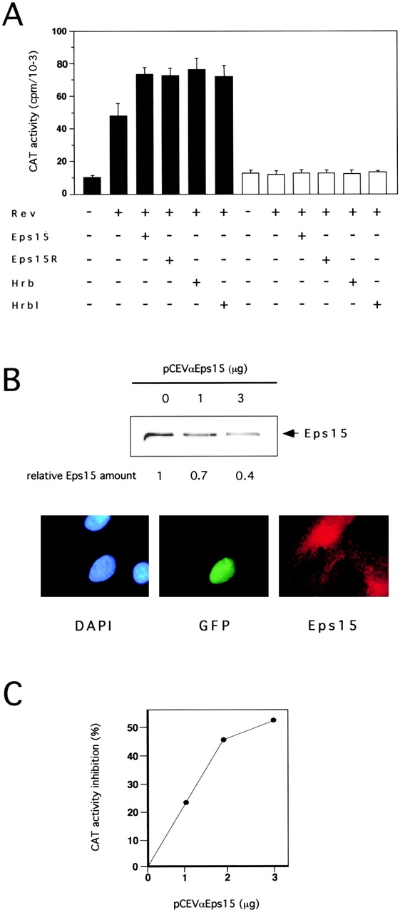
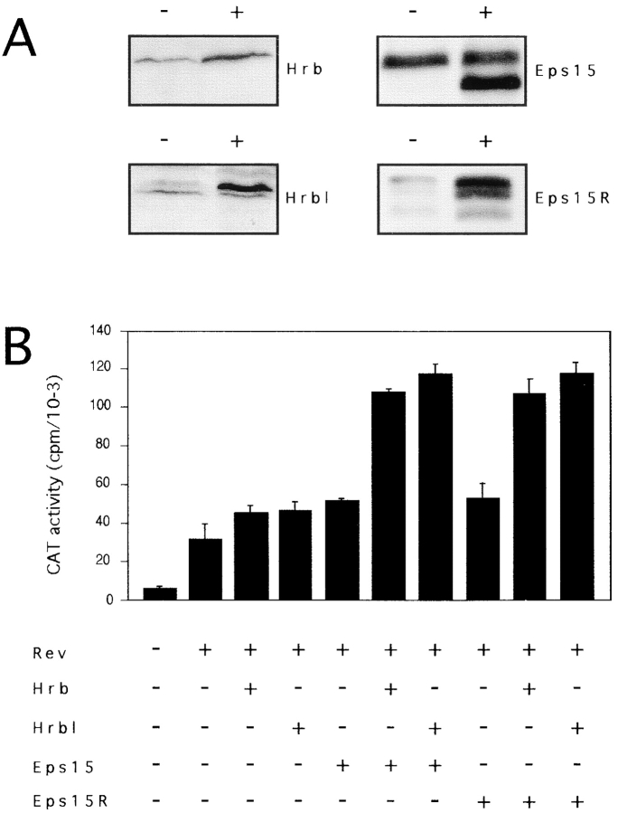
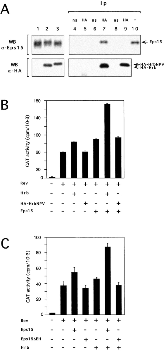
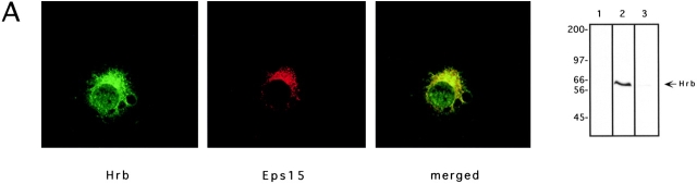
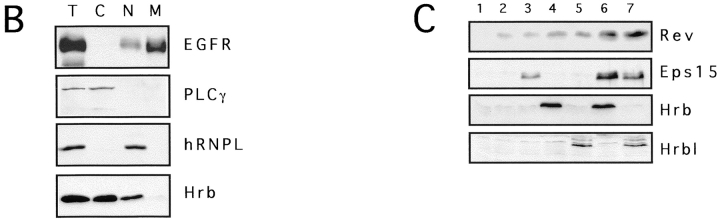
The Eps15 homology (EH) module is a protein-protein interaction domain that establishes a network of connections involved in various aspects of endocytosis and sorting. The finding that EH-containing proteins bind to Hrb (a cellular cofactor of the Rev protein) and to the related protein Hrbl raised the possibility that the EH network might also influence the so-called Rev export pathway, which mediates nucleocytoplasmic transfer of proteins and RNAs. In this study, we demonstrate that Eps15 and Eps15R, two EH-containing proteins, synergize with Hrb and Hrbl to enhance the function of Rev in the export pathway. In addition, the EH-mediated association between Eps15 and Hrb is required for the synergistic effect. The interaction between Eps15 and Hrb occurs in the cytoplasm, thus pointing to an unexpected site of action of Hrb, and to a possible role of the Eps15-Hrb complex in regulating the stability of Rev.


Figures

Figure 1
Figure 1
Eps15 and Eps15R influence Rev activity. (A) Rev-dependent CAT activity in cells transfected with the indicated vectors (underneath) and either the CAT reporter plasmid pDM128 (filled bars) or the control pDM138 (open bars). In this and all subsequent experiments, data are representative of at least three independent experiments, performed on individually transfected triplicates. (B, top) CV1 cells were transfected with the indicated amounts of pCEVαEps15. Cellular lysates (100 μg) were immunoblotted with an anti-Eps15 antibody and with an anti-tubulin antibody as a control (not shown). The levels of Eps15 protein, determined by densitometry and adjusted to account for variations in the level of tubulin, are indicated relative to the value in the mock-transfected lysate. (Bottom) CV1 cells were transfected with 0.5 μg of pNLS-GFP (as internal reference) and 5 μg of pCEVαEps15. Staining of cells was with rabbit anti-Eps15 antibody followed by CY3-conjugated anti-rabbit IgG and by nuclear counterstaining with DAPI. Photographs of the same field were taken with filters specific for DAPI-, GFP-, and Eps15(CY3)-specific fluorescences. The vast majority of cells expressing nuclear GFP, presumably expressing antisense Eps15 RNA, displayed a weaker signal for Eps15 as compared with untransfected cells. Cells transfected with pNLS-GFP alone or together with pCEV control vector showed normal levels of Eps15-specific staining (not shown). (C) CV1 cells were cotransfected with the pDM128 CAT reporter, Rev and with the indicated amounts of either pCEVαEps15 or pCEV empty vector. CAT activities are expressed as percent inhibition in pCEVαEps15 transfectants, compared with mock transfectants. In all above and subsequent experiments the total final amount of transfected DNA and the individual amounts of promoter units were kept constant, by adding appropriate amounts of control pDM, pMT, and pCEV empty vectors.
Figure 2
Figure 2
Eps15 and Eps15R synergize with Hrb and Hrbl. (A) Immunoblotting analysis of Eps15, Eps15R, Hrb, and Hrbl overexpression. Cellular proteins (100 μg) of CV-1 cells, transfected with expression vectors for Eps15, Eps15R, Hrb, and Hrbl (+ lanes) or with the corresponding empty vector (− lanes) were analyzed by immunoblotting with antibodies specific for each protein, as indicated. The overexpressed human Eps15 migrates as a faster band (140 kD) compared with the endogenous monkey protein (150 kD). In repeated experiments, we observed 3–10-fold overexpression of Eps15, Eps15R, Hrb or Hrbl when the corresponding expression vectors were transfected either alone of in combination. (B) Rev-dependent CAT activity in CV-1 cells transfected with pDM128 and the indicated expression vectors.
Figure 3
Figure 3
Interaction between Eps15 and Hrb is required for Rev activation. (A) C33A cells were transfected with 20 μg of pCEVEps15 together with 20 μg of either pMTHA-Hrb (lane 2) or pMTHA-HrbNPV (lane 3), or pMT control vector (lane 1). Cellular lysates were immunoblotted with an anti-Eps15 serum (top) or with an anti-HA antibody (bottom). We observed that, due to the presence of the four amino acid substitutions, HA-HrbNPV migrates as a faster band when compared with the corresponding HA-Hrb molecule. Cellular proteins from mock (lanes 4 and 5), HA-Hrb (lanes 6 and 7) or HA-HrbNPV (lanes 8 and 9) transfectants were immunoprecipitated with either an irrelevant monoclonal (ns-labeled lanes 4, 6, and 8) or with the anti-HA antibody (HA-labeled lanes 5, 7, and 9) and detected with an anti-Eps15 (top) or with the anti-HA (bottom) antibody. In the top panel, lane 10 corresponds to 50 μg of cellular lysate, to serve as a reference for positioning Eps15. (B and C) Rev-dependent CAT activity in CV-1 cells transfected with pDM128 and various mutants of Eps15 and Hrb (indicated underneath).
Figure 4
Figure 4
Eps15 and Hrb colocalize in the cytosol and increase Rev levels. (A) Subcellular localization of endogenous Eps15 and Hrb proteins in CV1 cells. Fluorescent cells were analyzed by optical scanning of horizontal sections using confocal microscopy. A representative horizontal section is shown (green, Hrb; red, Eps15; yellow, merged). The specificity of the anti-Hrb antibody was confirmed by immunoblot (right-most panel) on 100 μg of total proteins from CV-1 cells. Lane 1, staining with normal goat serum; lane 2, staining with anti-Hrb, lane 3, staining with anti-Hrb preincubated with the cognate peptide. Preincubation of the anti-Hrb antibody with the specific peptide also resulted in no signal in immunofluorescence analysis (not shown). (B) Subcellular fractionization. Subcellular fractions of CV-1 cells are identified as C (cytosolic), N (nuclear) and M (membrane), or T (total lysate). Aliquots of each fraction, representative of the same number of cells (1 × 10⁶), were analyzed by immunoblot for the presence of EGFR (membrane marker), PLC-γ (cytosolic marker), hRNPL (nuclear marker) and Hrb. As evident C and M fraction are >95% pure, whereas the nuclear fraction was ∼10% contaminated by membranes. (C) Steady state levels of the Rev protein in various transfectants. Immunoblot analysis with the antibodies indicated on the right was performed on CV-1 cells (2.5 × 10⁵) transfected with various combinations of plasmids. Transfectants were: 1, mock; 2, Rev; 3, Rev and Eps15; 4, Rev and Hrb; 5, Rev and Hrbl; 6, Rev, Eps15, and Hrb; 7, Rev, Eps15, and Hrbl.
Figure 4
Figure 4
Eps15 and Hrb colocalize in the cytosol and increase Rev levels. (A) Subcellular localization of endogenous Eps15 and Hrb proteins in CV1 cells. Fluorescent cells were analyzed by optical scanning of horizontal sections using confocal microscopy. A representative horizontal section is shown (green, Hrb; red, Eps15; yellow, merged). The specificity of the anti-Hrb antibody was confirmed by immunoblot (right-most panel) on 100 μg of total proteins from CV-1 cells. Lane 1, staining with normal goat serum; lane 2, staining with anti-Hrb, lane 3, staining with anti-Hrb preincubated with the cognate peptide. Preincubation of the anti-Hrb antibody with the specific peptide also resulted in no signal in immunofluorescence analysis (not shown). (B) Subcellular fractionization. Subcellular fractions of CV-1 cells are identified as C (cytosolic), N (nuclear) and M (membrane), or T (total lysate). Aliquots of each fraction, representative of the same number of cells (1 × 10⁶), were analyzed by immunoblot for the presence of EGFR (membrane marker), PLC-γ (cytosolic marker), hRNPL (nuclear marker) and Hrb. As evident C and M fraction are >95% pure, whereas the nuclear fraction was ∼10% contaminated by membranes. (C) Steady state levels of the Rev protein in various transfectants. Immunoblot analysis with the antibodies indicated on the right was performed on CV-1 cells (2.5 × 10⁵) transfected with various combinations of plasmids. Transfectants were: 1, mock; 2, Rev; 3, Rev and Eps15; 4, Rev and Hrb; 5, Rev and Hrbl; 6, Rev, Eps15, and Hrb; 7, Rev, Eps15, and Hrbl.

References

    1. Bogerd H.P., Fridell R.A., Madore S., Cullen B.R. Identification of a novel cellular cofactor for the Rev/Rex class of retroviral regulatory proteins. Cell. 1995;82:485–494. - PubMed
    1. Coda L., Salcini A.E., Confalonieri S., Pelicci G., Sorkina T., Sorkin A., Pelicci P.G., Di Fiore P.P. Eps15R is a tyrosine kinase substrate with characteristics of a docking protein possibly involved in coated pits-mediated internalization. J. Biol. Chem. 1998;273:3003–3012. - PubMed
    1. Cullen B.R. HIV-1 auxiliary proteinsmaking connections in a dying cell. Cell. 1998;93:685–692. - PubMed
    1. Fazioli F., Minichiello L., Matoskova B., Wong W.T., Di Fiore P.P. eps15, a novel tyrosine kinase substrate, exhibits transforming activity. Mol. Cell Biol. 1993;13:5814–5828. - PMC - PubMed
    1. Fischer U., Huber J., Boelens W.C., Mattaj I.W., Luhrmann R. The HIV-1 Rev activation domain is a nuclear export signal that accesses an export pathway used by specific cellular RNAs. Cell. 1995;82:475–483. - PubMed

Publication types