

Visualization of head-head interactions in the inhibited state of smooth muscle myosin
- PMID: 10613897
- PMCID: PMC2174251
- DOI: 10.1083/jcb.147.7.1385
Visualization of head-head interactions in the inhibited state of smooth muscle myosin
Abstract
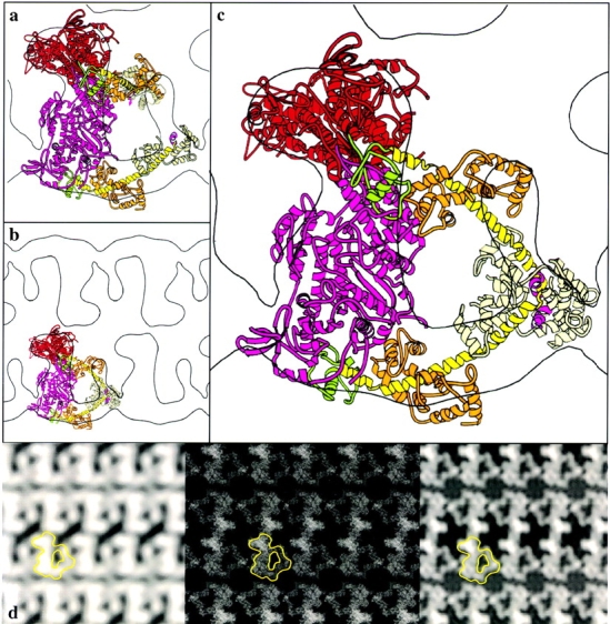
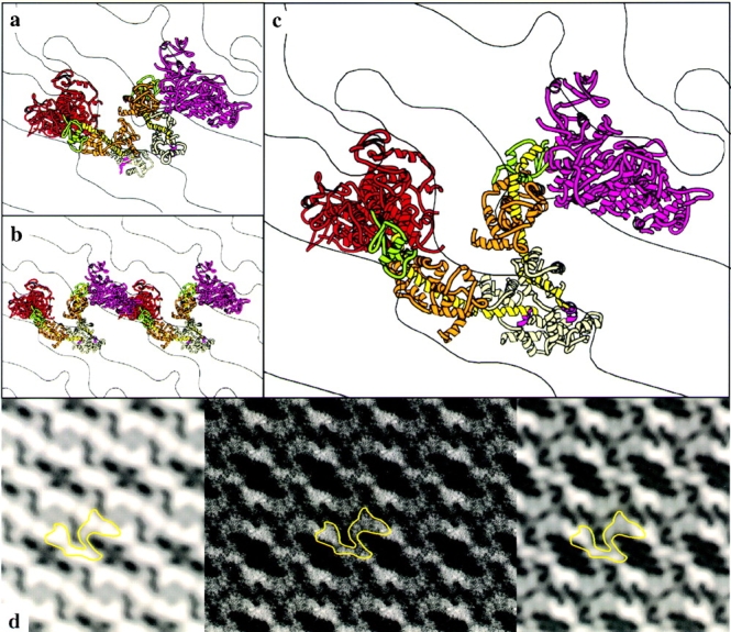
The structural basis for the phosphoryla- tion-dependent regulation of smooth muscle myosin ATPase activity was investigated by forming two- dimensional (2-D) crystalline arrays of expressed unphosphorylated and thiophosphorylated smooth muscle heavy meromyosin (HMM) on positively charged lipid monolayers. A comparison of averaged 2-D projections of both forms at 2.3-nm resolution reveals distinct structural differences. In the active, thiophosphorylated form, the two heads of HMM interact intermolecularly with adjacent molecules. In the unphosphorylated or inhibited state, intramolecular interactions position the actin-binding interface of one head onto the converter domain of the second head, thus providing a mechanism whereby the activity of both heads could be inhibited.
Figures
References
-
- Chiu W., Avila-Sakar A.J., Schmid M.F. Electron crystallography of macromolecular periodic arrays on phospholipid monolayers. Adv. Biophys. 1997;34:161–172. - PubMed
-
- Collaborative Computational Project, Number 4. 1994. The CCP4 suite: programs for protein crystallography. Acta Cryst. D50:760–763. - PubMed
-
- Craig R., Szent-Györgyi A.G., Beese L., Flicker P., Vibert P., Cohen C. Electron microscopy of thin filaments decorated with a Ca2+-regulated myosin. J. Mol. Biol. 1980;140:35–55. - PubMed
-
- Cremo C.R., Sellers J.R., Facemyer K.C. Two heads are required for phosphorylation-dependent regulation of smooth muscle myosin. J. Biol. Chem. 1995;270:2171–2175. - PubMed
-
- Crowther R.A., Henderson R., Smith J.M. MRC image processing programs. J. Struct. Biol. 1996;116:9–16. - PubMed
Publication types
MeSH terms
Substances
Grants and funding
LinkOut - more resources
Full Text Sources
Molecular Biology Databases
