. 1999 Dec 27;147(7):1481-92.
doi: 10.1083/jcb.147.7.1481.

Alternative splicing regulates the subcellular localization of A-kinase anchoring protein 18 isoforms

Affiliations

Alternative splicing regulates the subcellular localization of A-kinase anchoring protein 18 isoforms

K W Trotter et al. J Cell Biol. .

Abstract

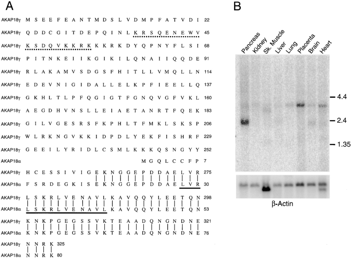
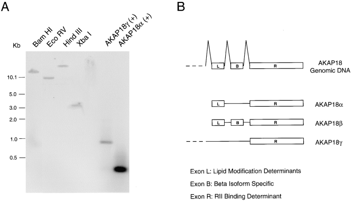
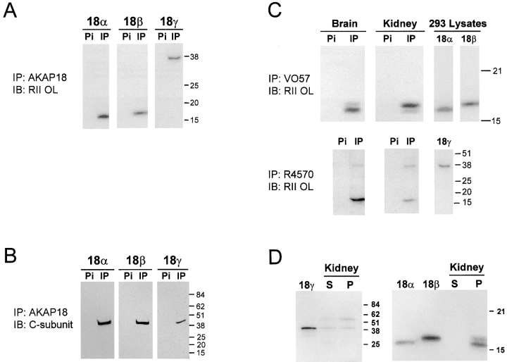
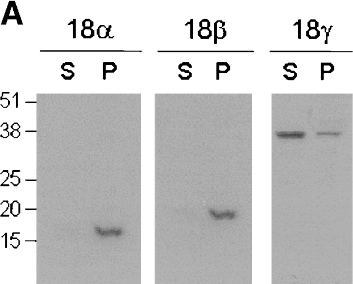
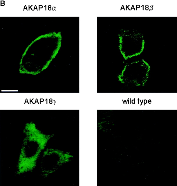
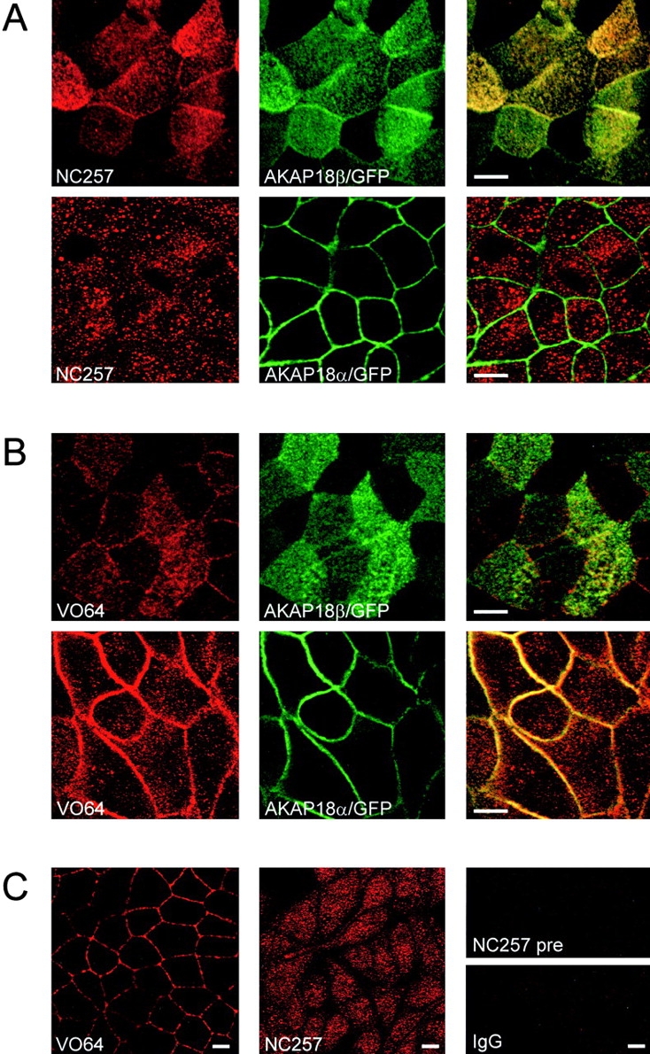
The cAMP-dependent protein kinase (PKA) is localized to specific subcellular compartments by association with A-kinase anchoring proteins (AKAPs). AKAPs are a family of functionally related proteins that bind the regulatory (R) subunit of PKA with high affinity and target the kinase to specific subcellular organelles. Recently, AKAP18, a low molecular weight plasma membrane AKAP that facilitates PKA-mediated phosphorylation of the L-type Ca(2+) channel, was cloned. We now report the cloning of two additional isoforms of AKAP18, which we have designated AKAP18beta and AKAP18gamma, that arise from alternative mRNA splicing. The AKAP18 isoforms share a common R subunit binding site, but have distinct targeting domains. The original AKAP18 (renamed AKAP18alpha) and AKAP18beta target the plasma membrane when expressed in HEK-293 cells, while AKAP18gamma is cytosolic. When expressed in epithelial cells, AKAP18alpha is targeted to lateral membranes, whereas AKAP18beta is accumulated at the apical membrane. A 23-amino acid insert, following the plasma membrane targeting domain, facilitates the association of AKAP18beta with the apical membrane. The data suggest that AKAP18 isoforms are differentially targeted to modulate distinct intracellular signaling events. Furthermore, the data suggest that plasma membrane AKAPs may be targeted to subdomains of the cell surface, adding additional specificity in intracellular signaling.


Figures

Figure 1
Figure 1
Cloning of human AKAP18γ. A, Comparison of human AKAP18γ and AKAP18α amino acid sequences. Common residues are indicated by a vertical line, a dashed line marks the putative nuclear localization signal, and the RII binding site is underlined. B, A human multiple tissue Northern blot was hybridized with a random primed 32P-labeled probe generated against the unique region of AKAP18γ (nt 357–689). The blot was stripped and rehybridized with a β-actin probe. mRNA size markers are shown in kb. Similar results were obtained in two separate blots.
Figure 2
Figure 2
Cloning of AKAP18β. A, Primers used to amplify human pancreas cDNA are shown schematically; PCR was performed using human pancreas cDNA and the primer pairs indicated. + indicates addition of 1 ng cDNA and − indicates no cDNA. Samples were electrophoresed on 1% agarose gels and visualized with ethidium bromide. mRNA size markers are shown in kb. No PCR product was observed using primers A + D, indicating that cDNAs containing both AKAP18γ- and -α–specific sequence does not exist in this tissue. The data are representative of three PCR reactions using human pancreas or lung cDNA as template. B, RT-PCR with AKAP18α-specific primers (C + B) was performed using cDNA from cultured cell lines, human pancreas, and human brain. cDNA quality was verified by amplifying each sample with human cyclophillin primers. + indicates AKAP18α or cyclophilin plasmid control and – indicates no addition of template. Samples were electrophoresed on 1.0% agarose gels and visualized with ethidium bromide. DNA size markers are shown in kb. Data are representative of two separate experiments. C, Comparison of AKAP18α and AKAP18β amino acid sequences. Common residues are indicated by a vertical line and the RII binding site is underlined. D, Schematic of three AKAP18 isoforms. The proteins are drawn to scale and unique regions marked by different shadings.
Figure 3
Figure 3
Alternative splicing gives rise to three distinct AKAP18 mRNAs. A, Human genomic DNA digested with the enzymes indicated, or positive control cDNAs were hybridized with a radiolabeled probe overlapping the common region of all three AKAP18 isoforms. DNA size standards are shown in kb. B, Schematic of the mouse AKAP18 gene and mRNAs encoding each AKAP18 isoform. Exons encoding the lipid modification determinant (Exon L), the AKAP18β-specific sequence (Exon B), and the RII binding region (Exon R) have been identified.
Figure 4
Figure 4
Analysis of AKAP18-related proteins. A, Lysates prepared from HEK-293 cells transiently expressing individual AKAP18 isoforms were immunoprecipitated with antisera directed against AKAP18. Samples were separated by SDS-PAGE and proteins visualized by RII overlay. B, Lysates prepared from HEK-293 cells transiently expressing individual AKAP18 isoforms were immunoprecipitated with rabbit antisera directed against AKAP18. Samples were separated by SDS-PAGE and blots were probed with mouse anti-PKA C subunit. C, Whole rat brain or kidney lysates were immunoprecipitated with rabbit antisera directed against AKAP18 as indicated. Samples were electrophoresed on SDS-PAGE and visualized by RII overlay. D, Whole rat kidney lysates were fractionated into soluble (S) and particulate (P) fractions in hypotonic buffers lacking detergent. Equal ratios of the soluble and particulate fractions were electrophoresed on SDS-PAGE and visualized by RII overlay. For C and D, lysates were also prepared from HEK-293 cells transiently transfected with AKAP18 isoforms, and samples were electrophoresed and analyzed in parallel with the tissue samples. For all panels, protein size standards are shown in kD and the data are representative of at least four similar experiments. IP, immunoprecipitation; IB, immunoblotting; Pi, preimmune sera.
Figure 5
Figure 5
Targeting of AKAP18 isoforms in HEK-293 cells. A, HEK-293 cells were transiently transfected with cDNAs encoding each AKAP18 isoform. Cells were separated into soluble (S) and particulate (P) fractions and 25 μg of each sample was resolved by SDS-PAGE. The distribution of AKAP18 isoforms was determined by immunoblot analysis using rabbit antisera directed against AKAP18α. Protein size standards are shown in kD. The data are representative of three similar experiments. B, HEK-293 cells were transiently transfected with AKAP18 cDNAs and plated on glass coverslips. Eight hours after transfection, wild-type and transfected cells were stained with affinity-purified rabbit anti-AKAP18 IgG (VO57; 1 μg/ml) and FITC-conjugated secondary antibody. The cells imaged are representative of at least four independent transfection studies with each plasmid. Bar, 10 μm.
Figure 5
Figure 5
Targeting of AKAP18 isoforms in HEK-293 cells. A, HEK-293 cells were transiently transfected with cDNAs encoding each AKAP18 isoform. Cells were separated into soluble (S) and particulate (P) fractions and 25 μg of each sample was resolved by SDS-PAGE. The distribution of AKAP18 isoforms was determined by immunoblot analysis using rabbit antisera directed against AKAP18α. Protein size standards are shown in kD. The data are representative of three similar experiments. B, HEK-293 cells were transiently transfected with AKAP18 cDNAs and plated on glass coverslips. Eight hours after transfection, wild-type and transfected cells were stained with affinity-purified rabbit anti-AKAP18 IgG (VO57; 1 μg/ml) and FITC-conjugated secondary antibody. The cells imaged are representative of at least four independent transfection studies with each plasmid. Bar, 10 μm.
Figure 6
Figure 6
AKAP18α and -β are differentially targeted in MDCK cells. A, MDCK cells stably expressing AKAP18α/GFP, AKAP18β/GFP, or GFP alone were grown on Transwell filters. Confluent monolayers were fixed in 4% paraformaldehyde and analyzed by confocal microscopy in XY and XZ planes. At least three individual cell lines expressing each construct were analyzed and similar results were obtained. Bar, 10 μm. B, The distributions of AKAP18α/GFP and AKAP18β/GFP were compared to the distribution of markers for tight junctions (ZO-1), adherens junctions (β-catenin), and apical membranes (gp135). Stably transfected cells were fixed and stained with rat anti ZO-1 (1:400), rabbit anti–β-catenin (1:400), or mouse anti-gp135 (1:50), followed by the appropriate Texas red-conjugated secondary antibody. Images are shown as XZ scans and are representative of images collected in two independent experiments.
Figure 7
Figure 7
Localization of endogenous AKAP18 in MDCK cells. A, MDCK cells stably expressing AKAP18β/GFP or AKAP18α/GFP were grown on Transwell filters and confluent monolayers were fixed in 4% paraformaldehyde. Cells were permeabilized, blocked, and stained with NC257 (1:1,000 dilution) followed by Texas red-conjugated secondary antibody. Antibody staining was compared with the distribution of the GFP fusion proteins by confocal microscopy. B, MDCK cells stably expressing AKAP18β/GFP or AKAP18α/GFP were grown on Transwell filters and confluent monolayers were fixed and stained with VO64 (1 μg/ml) as described in A. For experiments in A and B, scanning in one channel was performed with the other laser off to assure that there was no bleed-through. C, Wild-type MDCK cells were grown on Transwell filters and confluent monolayers were fixed and stained with VO64 or NC257 as described above. Preimmune sera and normal rabbit IgG at the same concentrations failed to stain any structures in wild-type or transfected MDCK cells. Bar, 10 μm.
Figure 8
Figure 8
The unique 23 amino acids in AKAP18β facilitate apical targeting. A, Schematic diagram of the three GFP fusion proteins expressed in HEK-293 and MDCK cells. GFP is not drawn to scale. The unique region of AKAP18β is filled in black. B, Each construct was expressed transiently in HEK-293 cells and the distributions of the expressed GFP-tagged proteins analyzed by confocal microscopy. Bar, 10 μm. The images are representative of three individual experiments. C, MDCK cells stably expressing the constructs shown in A were grown to confluence on glass coverslips and the distribution of the GFP chimeras was determined by confocal microscopy. Images were collected in the XY and XZ planes. Similar results were obtained in transient transfection assays and in at least three clonal cell lines for each construct. Bar, 10 μm.

References

    1. Ali S., Chen X., Lu M., Xu J.Z., Lerea K.M., Hebert S.C., Wang W.H. The A kinase anchoring protein is required for mediating the effect of protein kinase A on ROMK1 channels. Proc. Natl. Acad. Sci. USA. 1998;95:10274–10278. - PMC - PubMed
    1. Angelo R., Rubin C.S. Molecular characterization of an anchor protein (AKAPCE) that binds the RI subunit (RCE) of type I protein kinase A from Caenorhabditis elegans . J. Biol. Chem. 1998;273:14633–14643. - PubMed
    1. Brown D.A., London E. Functions of lipid rafts in biological membranes. Annu. Rev. Cell Dev. Biol. 1998;14:111–136. - PubMed
    1. Burton K.A., Johnson B.D., Hausken Z.E., Westenbroek R.E., Idzerda R.L., Scheuer T., Scott J.D., Catterall W.A., McKnight G.S. Type II regulatory subunits are not required for the anchoring-dependent modulation of Ca2+ channel activity by cAMP-dependent protein kinase. Proc. Natl. Acad. Sci. USA. 1997;94:11067–11072. - PMC - PubMed
    1. Carr D.W., Stofko-Hahn R.E., Fraser I.D., Bishop S.M., Acott T.S., Brennan R.G., Scott J.D. Interaction of the regulatory subunit (RII) of cAMP-dependent protein kinase with RII-anchoring proteins occurs through an amphipathic helix binding motif. J. Biol. Chem. 1991;266:14188–14192. - PubMed

Publication types

MeSH terms

Associated data