. 1999 Dec 27;147(7):1533-48.
doi: 10.1083/jcb.147.7.1533.

Ezrin promotes morphogenesis of apical microvilli and basal infoldings in retinal pigment epithelium

Affiliations

Ezrin promotes morphogenesis of apical microvilli and basal infoldings in retinal pigment epithelium

V L Bonilha et al. J Cell Biol. .

Abstract

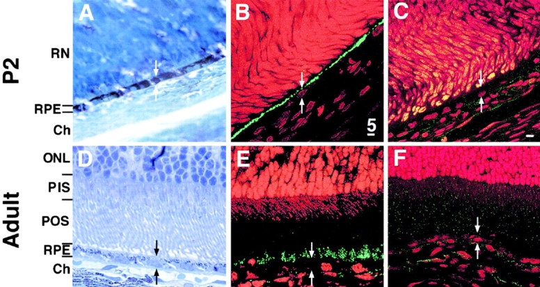
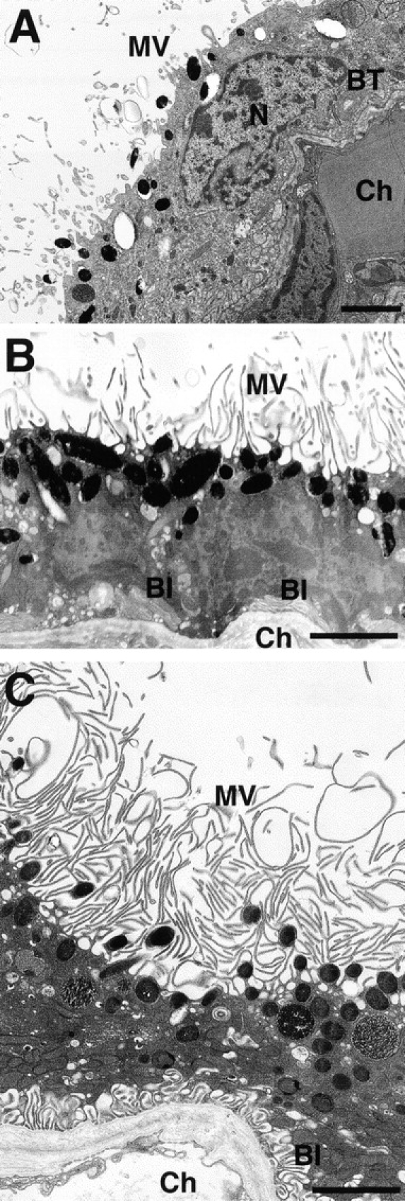
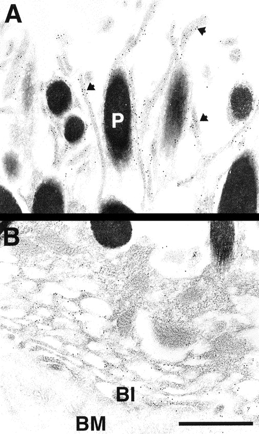
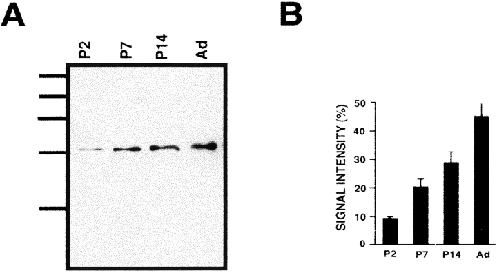
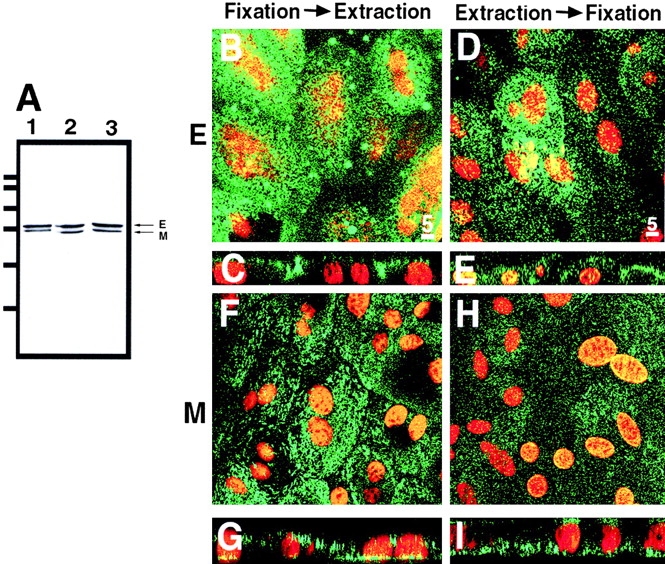
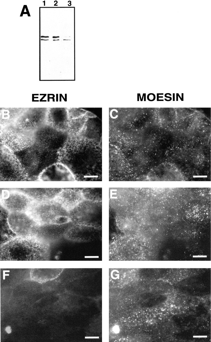
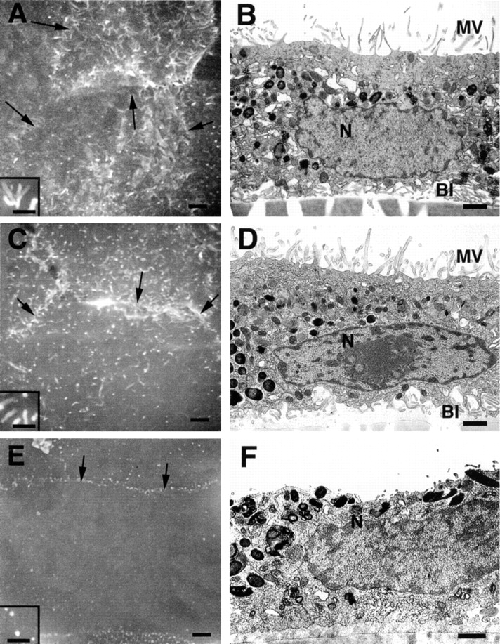
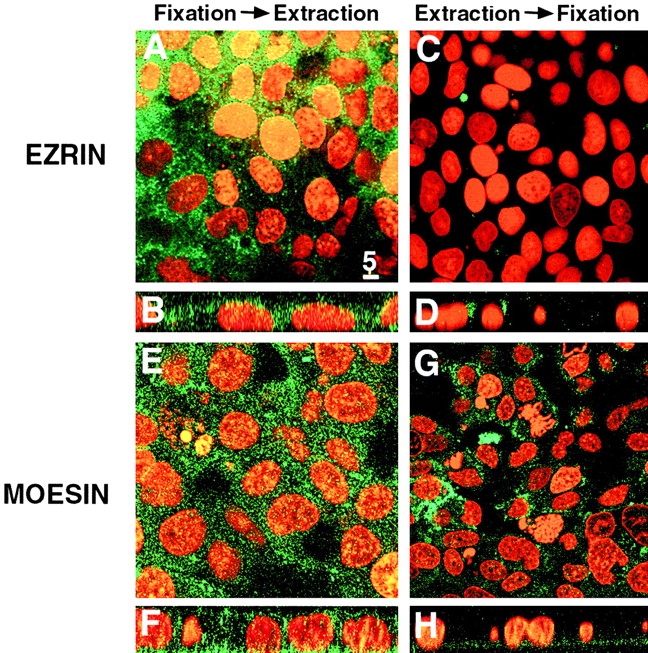
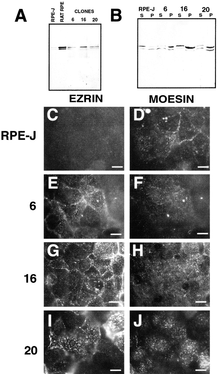
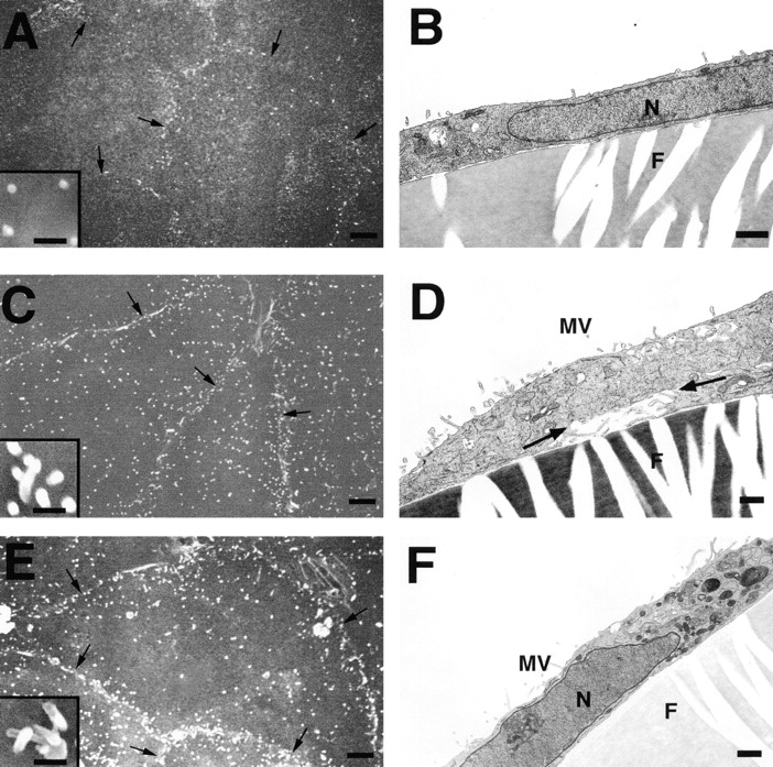
Ezrin, a member of the ezrin/radixin/moesin (ERM) family, localizes to microvilli of epithelia in vivo, where it bridges actin filaments and plasma membrane proteins. Here, we demonstrate two specific morphogenetic roles of ezrin in the retinal pigment epithelium (RPE), i.e., the formation of very long apical microvilli and of elaborate basal infoldings typical of these cells, and characterize the role of ezrin in these processes using antisense and transfection approaches. In the adult rat RPE, only ezrin (no moesin or radixin) was detected at high levels by immunofluorescence and immunoelectron microscopy at microvilli and basal infoldings. At the time when these morphological differentiations develop, in the first two weeks after birth, ezrin levels increased fourfold to adult levels. Addition of ezrin antisense oligonucleotides to primary cultures of rat RPE drastically decreased both apical microvilli and basal infoldings. Transfection of ezrin cDNA into the RPE-J cell line, which has only trace amounts of ezrin and moesin, sparse and stubby apical microvilli, and no basal infoldings, induced maturation of microvilli and the formation of basal infoldings without changing moesin expression levels. Taken together, the results indicate that ezrin is a major determinant in the maturation of surface differentiations of RPE independently of other ERM family members.


Figures

Figure 1
Figure 1
Ezrin localizes to the apical surface of rat RPE during postnatal maturation. RPE cells undergo a dramatic change in their morphology during postnatal maturation of the retina, which also includes the formation of PR outer segments. Observation of thick sections of epon-embedded immature (P2, A) and mature (Adult, D) eyes stained with Toluidine blue highlights the cell layers present in each situation. Immature RPE cells are very close apically to the immature retinal nuclei (RN) and interact basally with the choroid (Ch). On the other hand, fully differentiated RPE cells interact apically with the PR outer segments (POS). For ezrin immunolocalization, tissues were fixed by intracardiac perfusion with 4% paraformaldehyde. 10-μm cryosections of eyecups were labeled with a mAb to ezrin (B and E), while parallel sections were stained with a pAb directed to moesin (C and F). Cell nuclei were labeled with propidium iodide. The labeled cryosections were analyzed by dual channel laser scanning confocal microscopy. Immature (B) and mature (E) RPE cells displayed ezrin localization almost exclusively at the apical RPE surface. Neither immature (C) nor mature (F) RPE cells display any moesin staining. Moesin staining was detected in the underlying choroid. Ch, choroid; ONL, outer nuclear layer; PIS, PR inner segments; POS, PR outer segments; RN, immature retinal nuclei. Bar, 5 μm.
Figure 2
Figure 2
Ultrastructural maturation of the retinal pigment epithelium. Neural retina-free eyecups were fixed in 2.5% glutaraldehyde + 0.2% picric acid made in 0.1 M cacodylate buffer, and processed for transmission EM. A, The newborn RPE (P2) has very short microvilli extending from its apical surface. Its basal surface interacts with the choriocapillaris through the relatively smooth Bruch's basement membrane. Cytoplasmic tubular structures (BT) can be observed connected to the basal surface. B, At P7, longer microvilli emerge from the apical surface. The basal surface displays deep basal infoldings in some areas. C, In the mature rat eye, RPE cells extend remarkably thin and long microvilli from their apical surfaces. BI, basal infoldings; BT, basal tubules; Ch, choroid; MV, microvilli; N, nucleus. Bars, 2 μm.
Figure 3
Figure 3
Ezrin localizes both at RPE apical microvilli and basal infoldings. Adult neural retina-free eyecups were fixed in a mix of 4% paraformaldehyde, 0.1% glutaraldehyde, and 0.2% picric acid prepared in PHEM modified buffer. Tissue was sequentially dehydrated in ethanol, embedded in Unicryl, and polymerized at −20°C under UV light. Ultrathin sections were sequentially reacted with a polyclonal antiezrin antibody and a gold-conjugated (10 nm) donkey anti–rabbit antibody. In these samples, labeling was specifically associated with apical microvilli (A) and to a lower extent with basal infoldings (B). Arrowheads point to colloidal gold/ezrin antibodies decorating microvilli. BI, basal infoldings; BM, Bruch's membrane; P, pigment granule. Bar, 0.5 μm.
Figure 4
Figure 4
Ezrin expression increases during postnatal maturation of RPE. A, RPE of different ages (P2 to adult [Ad]) were harvested and lysed in 2% SDS in PBS supplemented with protease inhibitors and 100 μM sodium orthovanadate. 10 μg of protein of each sample was separated in a 7.5% SDS gel, transferred to nitrocellulose membranes and probed with a pAb specific to a COOH-terminal peptide common to all ERM proteins, followed by ECF detection of immunoreactivity. B, Membranes were exposed to film and ezrin signal intensities were analyzed with NIH Image 1.62 software and plotted. During RPE maturation, ezrin expression is upregulated fourfold. Data shown are the mean of four independent experiments ± SEM. The large error bar associated with the adult samples is due to apical surface damage during the enzymatic peeling of the neural retina from the RPE.
Figure 5
Figure 5
Ezrin localizes mostly to the apical surface of primary RPE cells whereas moesin localizes mostly to the basal surface. Primary RPE monolayers were probed with an antibody specific to a COOH-terminal peptide conserved in all three ERM proteins. Whole extracts (10 μg) of these cells revealed the presence of two bands corresponding to the molecular weight of ezrin and moesin (A, lane 1, arrows), but no band corresponding to radixin, with intermediate mobility between the two other proteins. Analysis of Triton-extracted fractions from primary RPE cultures (20–30 μg and ∼60–80 μg protein per lane in the detergent soluble and insoluble fractions, respectively) showed that 45 ± 0.5% of ezrin and 52 ± 3.5% of moesin were extracted (A, lanes 2 [soluble] and 3 [insoluble]). Values reported are the mean of three independent experiments ± SEM. Differentiated monolayers plated on filters were fixed with paraformaldehyde, permeabilized with Triton X-100 for 10 min, and labeled either with an mAb to ezrin (E; B–E) or a pAb to moesin (M; F–I). Nuclei were stained with propidium iodide. Samples were observed using a dual channel laser scanning confocal microscope. B, Paraformaldehyde-fixed monolayers grown on polycarbonate filters displayed a punctate ezrin staining suggestive of an apical localization in primary RPE cultures. C, The apical localization of ezrin was confirmed in Z-scans performed through the monolayer. D and E, Confocal immunofluorescence analysis of primary RPE cultures extracted with 0.5% Triton X-100 and fixed with paraformaldehyde, revealed the presence of ezrin, primarily in apical microvilli, indicating that ezrin resisted nonionic detergent extraction mostly at the apical surface of the cells. F, On the other hand, moesin was more evenly distributed in the primary cells. G, Vertical section of the monolayers confirmed moesin distribution in the apical, lateral, and basal surface of primary RPE cells. H, Observation of cells extracted with 0.5% Triton and then fixed with paraformaldehyde, revealed that there was a decrease of moesin staining corresponding to moesin detergent extraction. I, Cross-sections through the monolayers revealed that the majority of moesin resistant to detergent extraction is localized to the basal surface of the cells suggesting that in these cells, moesin is weakly or not anchored to the apical cytoskeleton. Bar, 5 μm.
Figure 6
Figure 6
Treatment of primary RPE monolayers with ezrin oligonucleotides decreases ezrin expression and disrupts RPE microvilli. Immunoblot and immunofluorescence. Primary cultures of rat RPE were plated on filters. Cultures were supplemented with 20 μM sense or antisense oligonucleotides directed to ezrin for 96 h. At the end of this period, filters were cut in two pieces and processed separately. A, Half of filters were lysed in 2% PBS plus protease and phosphatase inhibitors and were biochemically analyzed. Antisense treated monolayers (lane 3) showed a 80% decrease in ezrin levels when compared with control (lane 1) and sense (lane 2) treated monolayers. The other half of the samples were permeabilized with 0.02% Triton X-100 for 10 min, fixed with paraformaldehyde, and processed for double immunofluorescence for ezrin and moesin. Samples were observed using an epifluorescence microscope, and images were collected with a cooled CCD camera. Digitally acquired images were processed using the Metamorph software. B, Primary cultures of RPE from mature eyes supplemented with buffer showed a typical microvilli staining pattern for ezrin. C, Moesin expression was diffuse throughout the cytoplasm of the cells with puncta of undefined origin. No changes in the ezrin (D) and moesin (E) labeling were observed when monolayers were supplemented with ezrin sense oligonucleotides. In sharp contrast, monolayers supplemented with antisense oligonucleotides almost completely lost their ezrin staining (F) while their moesin staining (G) did not change significantly from the control samples. Bars, 10 μm.
Figure 7
Figure 7
Treatment of primary cultures with ezrin antisense oligonucleotide disrupts apical microvilli and basal infoldings. Scanning and transmission EM. Primary RPE cultures plated on glass coverslips or on filters were treated with 20 μM sense or antisense oligonucleotides directed to ezrin for 96 h as previously described. After treatment, monolayers were fixed in 2.5% glutaraldehyde, sequentially dehydrated, and processed for observation at both scanning (A, C, and E) and transmission (B, D, and F) EM. Untreated primary RPE cultures (A and B) displayed an apical surface densely covered by microvilli and a basal surface with elaborated basal infoldings (B). Strikingly, monolayers treated with ezrin antisense oligonucleotide displayed a smooth surface, almost completely deprived of microvilli (E and F) and basal infoldings (F). On the other hand, monolayers treated with ezrin sense oligonucleotides displayed normal microvilli on their apical surfaces (C and D) and elaborated basal infoldings (D). Insets A, C, and E show that not only the number, but also the length, of microvilli is affected by the treatment of the cultures with ezrin antisense oligonucleotides. Adhesion sites between cells are indicated by the small arrows. BI, basal infoldings; MV, microvilli; N, nuclei. Bars: (A, C, and E) 2 μm; (insets in A, C, and E) 0.5 μm; (B, D and F) 1 μm.
Figure 8
Figure 8
Ezrin is mostly cytoplasmic, whereas moesin is mostly bound to the basolateral surface of RPE-J cells. Polarized RPE-J monolayers were plated on filters, processed as indicated below, and labeled either with an mAb specific to ezrin (A–D) or with a pAb specific to moesin (E–H). RPE-J cells fixed with paraformaldehyde before extraction with Triton X-100 displayed ezrin (A and B) and moesin (E and F) labeling diffusely distributed in the cytoplasm. Most of the ezrin staining was removed when cells were extracted before fixation (C and D). Moesin was detected mostly at the basal surface of cells extracted before fixation, suggesting preferential attachment to the basolateral cytoskeleton of RPE-J cells (G and H). Bar, 5 μm.
Figure 9
Figure 9
Overexpression of ezrin in RPE-J cells promotes its localization into detergent-resistant apical microvilli. A, 10 μg of whole cell lysates of RPE-J and primary cultures were loaded into 7.5% SDS-PAGE gels, and resolved proteins were transferred to nitrocellulose membranes, followed by immunoblot with a pAb to a peptide conserved in all three ERM proteins. Values shown are the mean of four independent experiments. Ezrin levels in wild-type RPE-J cells (RPE-J) were three times lower than in primary RPE cells (RAT RPE); ezrin:moesin ratio was 1.15:1 in RPE-J and 1.7 in primary RPE cultures. RPE-J clones overexpressing ezrin showed two times (clone 6) or three to four times (clones 16 and 20) higher total ezrin expression levels than wild-type RPE-J cells (RPE-J). B, RPE clones overexpressing ezrin displayed larger pools of ezrin associated with the cytoskeleton. In wild-type RPE-J cells (RPE-J, P), 37 ± 2.1% of ezrin and 57 ± 3.8% of moesin was resistant to extraction by Triton X-100. In RPE-J clones (6, 16, and 20, P), the cytoskeleton-associated fraction of ezrin was increased to 63 ± 8.6%, 58 ± 3.5%, and 69 ± 6.8% for clones 6, 16, and 20, respectively. Moesin partition into the detergent-resistant fraction was 53 ± 6.9%, 47 ± 5.6%, and 40 ± 7.3%, for clones 6, 16, and 20, respectively. 20–30 μg and ∼60–80 μg of the detergent soluble and insoluble fractions were loaded per lane. C–J, Immunofluorescence of RPE-J clones overexpressing ezrin. Wild-type RPE-J cells (C and D) and RPE-J clones overexpressing ezrin (E–J) were extracted with Triton X-100, fixed, and stained with an antibody against a VSVG epitope (C, E, G, and I) and an antibody against moesin (D, F, H, and J). Control RPE-J cells showed a low background staining with the VSVG epitope antibody (C). All three RPE-J clones overexpressing ezrin displayed a strong and homogeneous apical microvilli pattern that was resistant to detergent extraction (E, G, and I). Detergent-resistant moesin in RPE-J cells had a basolateral distribution (D), but acquired a more diffuse distribution and was present in puncta of undefined origin in RPE-J clones (F, H, and J). Bar, 10 μm.
Figure 10
Figure 10
RPE-J clones overexpressing ezrin develop apical microvilli-like structures and basal infoldings. The ultrastructure of RPE-J cells overexpressing ezrin was analyzed by transmission EM. Polarized monolayers plated on glass coverslips or on filters were fixed in 2.5% glutaraldehyde, sequentially dehydrated, and observed both by scanning (A, C, and E) and transmission (B, D, and F) EM. Untransfected RPE-J cells showed scattered stubby microvilli at their apical surface (A and B) and a smooth basal surface (B). Clones 6 and 20, overexpressing ezrin, developed numerous apical microvilli-like structures (C–F) and some basal infoldings (D, arrows). Insets in A, C, and E show that not only the number, but also the length, of microvilli is affected by the overexpression of exogenous ezrin in RPE-J cultures. Adhesion sites between cells are indicated by the small arrows. F, filter; MV, microvilli; N, nucleus. Bars: (A, C and E) 2 μm; (insets in A, C, and E) 0.5 μm; (B, D, and F) 1 μm.

References

    1. Algrain M., Turunen O., Vaheri A., Louvard D., Arpin M. Ezrin contains cytoskeleton and membrane binding domains accounting for its proposed role as a membrane–cytoskeletal linker. J. Cell Biol. 1993;120:129–139. - PMC - PubMed
    1. Amieva M.R., Litman P., Huang L., Ichimaru E., Furthmayr H. Disruption of dynamic cell surface architecture of NIH3T3 fibroblasts by the N-terminal domains of moesin and ezrinin vivo imaging with GFP fusion proteins. J. Cell Sci. 1998;112:111–125. - PubMed
    1. Andreoli C., Martin M., Le Borgne R., Reggio H., Mangeat P. Ezrin has properties to self-associate at the plasma membrane. J. Cell Sci. 1994;107:2509–2521. - PubMed
    1. Arpin M., Algrain M., Louvard D. Membrane-actin microfilament connectionsan increasing diversity of players related to band 4.1. Curr. Opin. Cell Biol. 1994;6:136–141. - PubMed
    1. Barila D., Murgia C., Nobili F., Perozzi G. Transcriptional regulation of the ezrin gene during rat intestinal development and epithelial differentiation. Biochim. Biophys. Acta. 1995;1263:133–140. - PubMed

Publication types

MeSH terms