. 2000 Jan;115(1):51-8.
doi: 10.1085/jgp.115.1.51.

The lipid-protein interface of a Shaker K(+) channel

Affiliations

The lipid-protein interface of a Shaker K(+) channel

K H Hong et al. J Gen Physiol. 2000 Jan.

Abstract

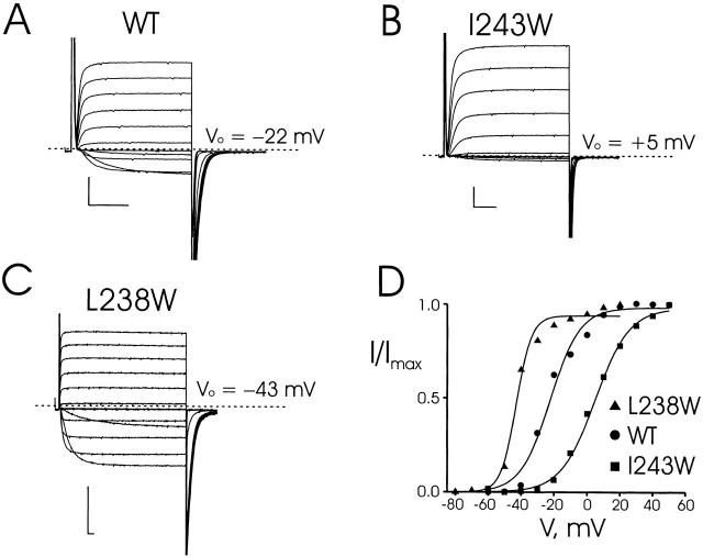
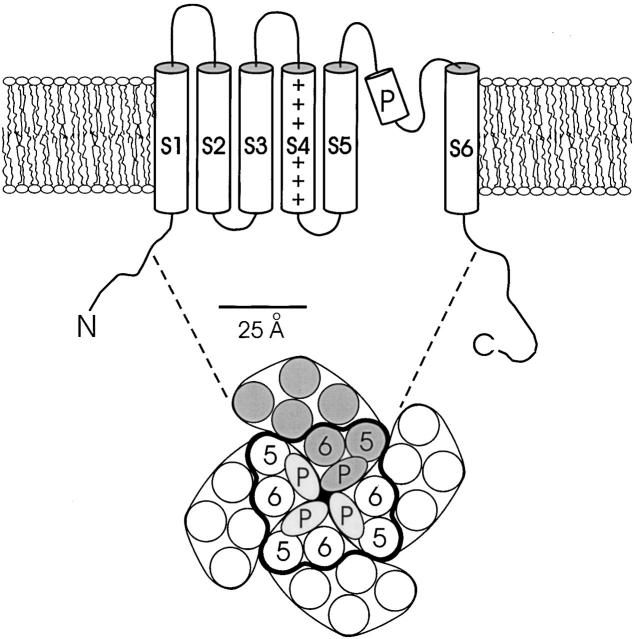
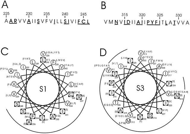
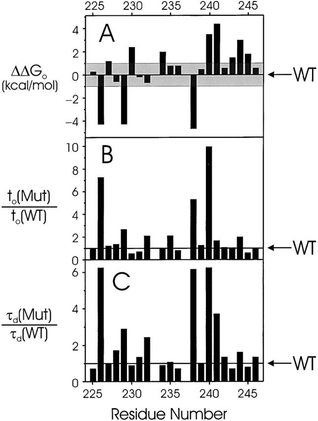
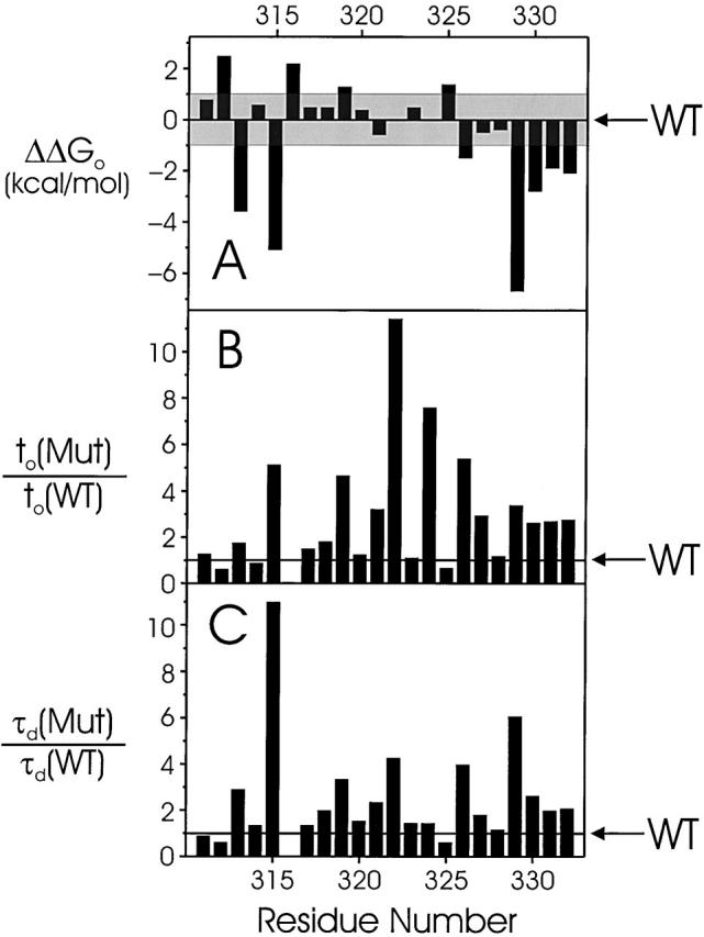
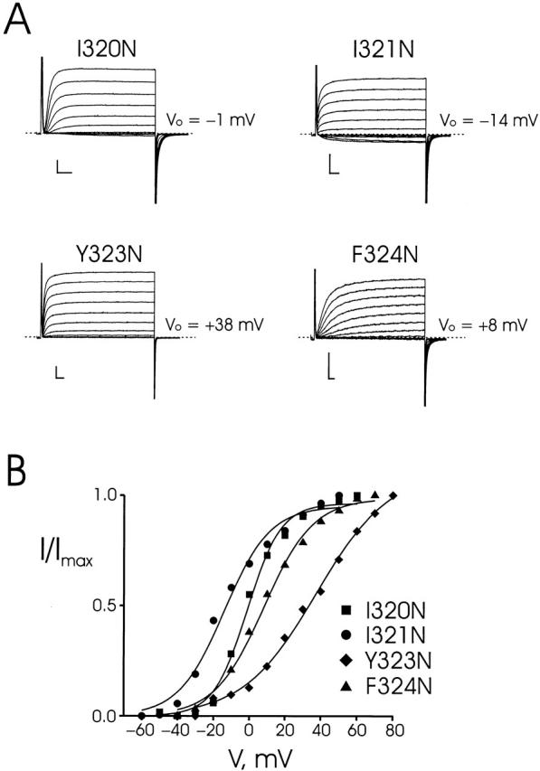
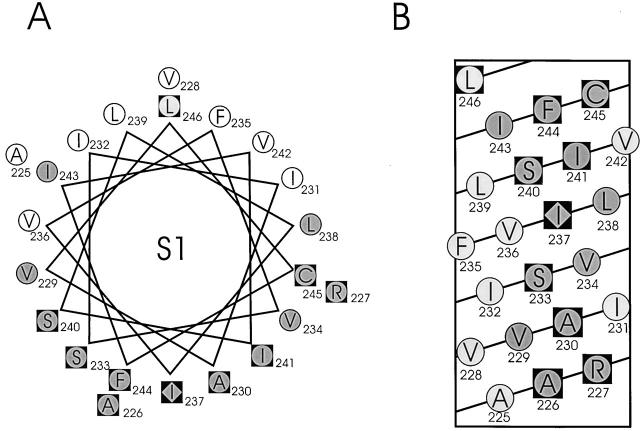
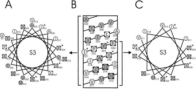
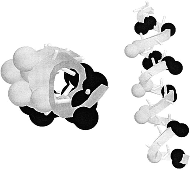
Tryptophan-substitution mutagenesis was applied to the first and third transmembrane segments (S1 and S3) of a Shaker-type K(+) channel for the purpose of ascertaining whether these sequences are alpha-helical. Point mutants were examined for significant functional changes, indicated by the voltage-activation curves and gating kinetics. Helical periodicity of functional alteration was observed throughout the entire S1 segment. A similar result was obtained with the first 14 residues of S3, but this periodicity disappeared towards the extracellular side of this transmembrane sequence. In both helical stretches, tryptophan-tolerant positions are clustered on approximately half the alpha-helix surface, as if the sidechains are exposed to the hydrocarbon region of the lipid bilayer. These results, combined with an analogous study of S2 (Monks, S., D.J. Needleman, and C. Miller. 1999. J. Gen. Physiol. 113:415-423), locate S1, S2, and S3 on the lipid-facing periphery of K(v) channels.


Figures

Figure 3
Figure 3
Recordings from tryptophan-substituted Shaker channels. (A) Two-electrode voltage-clamp recordings of wild-type channels expressed in Xenopus oocytes. Currents were recorded in KD98 medium with a holding potential of −90 mV, test pulses (30-ms duration) from −60 to +50 mV in 10-mV increments, and a tail potential of −70 mV. (B and C) Similar recordings taken for mutations I243W and L238W. For L238W, test pulses were from −80 to +20 mV with duration of 200 ms and each test pulse was followed by a fixed tail voltage −80 mV. (D) Voltage-activation curves for wild-type, I243W, and L238W mutant channels, calculated from tail-current analysis. Solid curves are Boltzmann fits to the equilibrium activation data. Scale bars in all data panels represent 1 μA, 10 ms.
Figure 1
Figure 1
Standard model for membrane topology of Kv channels. The lower cartoon represents an aerial view of the assembled tetramer from the extracellular side. The positions of S5, P-loop, and S6, encircled by heavy curve, are modeled to scale from a slice through the structure of the KcsA channel at the level of the external lipid headgroups. Helices are represented by circles of 11.8 Å diameter. Unlabeled helices represent hypothetical positions of S1–S4.
Figure 2
Figure 2
Sequence characteristics of S1 and S3 segments in Shaker. (A and B) Amino acid sequences of S1 and S3. Boldface underlined residues are those conserved in 120 S1 and 135 S3 K+ channel sequences, using a BLAST search (Altschul et al. 1997) of the nonredundant database. (C and D) α-helical projections of S1 and S3. Conserved residues, inscribed in black squares, are identified by two criteria. First, the most common residue is found in over 65% of instances; second, no more than four alternative substitutes are observed, and these must all be of similar chemical character. Listed in parentheses are alternative amino acids found at equivalent positions in other K+ channels.
Figure 4
Figure 4
Gating parameters of S1 tryptophan scan. Changes of empirical gating parameters resulting from point Trp mutations are displayed versus residue number. (A) Intrinsic free energy of open-state stabilization, ΔΔG o. The shaded region (|ΔΔG o| < 1 kcal/mol) represents the range of values defining tolerant residues. (B and C) Mutant-to-wild-type ratio of activation times, t o, and deactivation time constant, τd, determined as in materials and methods. Electrophysiological parameters are reported in Table .
Figure 5
Figure 5
Gating parameters of S3 tryptophan scan. Details are as in Fig. 4. Electrophysiological parameters are reported in Table .
Figure 6
Figure 6
Effects of Asn substitutions at certain Trp-tolerant positions. Two-electrode voltage clamp recordings (A) and voltage-activation curves (B) of the indicated mutants displayed as in Fig. 3. Electrophysiological parameters are reported in Table .
Figure 7
Figure 7
Results of Trp-scanning mutagenesis of S1. An α-helical wheel (A) and a net (B) diagram of S1 are shown with residues scored as either not expressed (diamonds), high impact (shaded circles), or tolerant (open circles), according to criteria described in text. Black squares represent conserved residues as in Fig. 2.
Figure 8
Figure 8
Results of Trp and Asn mutagenesis of S3. An α-helical wheel (A) and a net (B) diagram of S3 are shown as in Fig. 7. *Residues that were tested by Asn substitution. Striped circles represent ambiguous residues on the basis of Asn mutants, as discussed in text. (C) A restricted helical wheel diagram limited to the first 14 residues of S3.
Figure 9
Figure 9
Structural interpretation of sequence variability in S5. The first 16 residues in the outer helix of KcsA (positions 29–45), equivalent to S5 in Shaker (396–412), are represented in two views. (Left) View down the helix axis from the intracellular end; (right) longitudinal view, with intracellular end at bottom. (Black atoms) α and β carbons of sidechains packed directly against protein in KcsA; (grey atoms) α and β carbons of positions equivalent to variable residues in S5.

Comment in

References

    1. Altschul S.F., Madden T.L., Schaffer A.A., Zhang J., Zhang Z., Miller W., Lipman D. Gapped BLAST and PSI-BLASTa new generation of protein database search programs. Nucleic Acids Res. 1997;25:3389–3402. - PMC - PubMed
    1. Baldwin J.M., Schertler G.F.X., Unger V.M. An alpha-carbon template for the transmembrane helices in the rhodopsin family of G-protein–coupled receptors. J. Mol. Biol. 1997;272:144–164. - PubMed
    1. Choe S., Stevens C.F., Sullivan J.M. Three distinct structural environments of a transmembrane domain in the inwardly rectifying K+ channel ROMK1 defined by perturbation. Proc. Natl. Acad. Sci. USA. 1995;92:12046–12049. - PMC - PubMed
    1. Donnelly D., Overington J.P., Ruffle S.V., Nugent J.H., Blundell T.L. Modeling alpha-helical transmembrane domainsthe calculation and use of substitution tables for lipid-facing residues. Protein Sci. 1993;2:55–70. - PMC - PubMed
    1. Doyle D.A., Cabral J.M., Pfuetzner R.A., Kuo A., Gulbis J.M., Cohen S.L., Chait B.T., MacKinnon R. The structure of the K+ channelmolecular basis of K+ conduction and selectivity. Science. 1998;280:69–77. - PubMed

Publication types

Substances