

Identification and characterization of immunoglobulin G in blood as a major inhibitor of diagnostic PCR
- PMID: 10618113
- PMCID: PMC88721
- DOI: 10.1128/JCM.38.1.345-350.2000
Identification and characterization of immunoglobulin G in blood as a major inhibitor of diagnostic PCR
Abstract
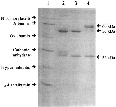
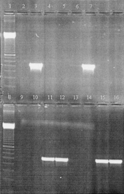
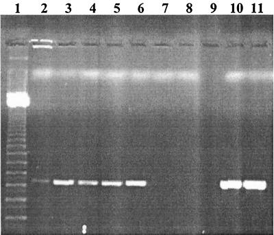
A major inhibitor of diagnostic PCR in human plasma was identified and the mechanism of inhibition was characterized. Human blood was divided by centrifugation into buffy coat, plasma, platelets, and erythrocytes. All these blood fractions were found to be highly inhibitory to a standardized PCR mixture containing the thermostable DNA polymerase AmpliTaq Gold. PCR inhibitors in human plasma were purified by chromatographic procedures and were characterized by a process of elimination, so that the PCR-inhibitory effects of plasma fractions were tested after each purification step. The major inhibitor in human plasma, as determined by size-exclusion chromatography, anion-exchange chromatography, and chromatofocusing, was found to be immunoglobulin G (IgG) on the basis of N-terminal amino acid sequencing and electrophoretic analysis of the purified polypeptide. When different concentrations of purified plasma IgG (PIgG) were added to PCR mixtures containing 11 different thermostable DNA polymerases and 1 ng of Listeria monocytogenes DNA as template DNA, the only polymerase that resisted inhibition was rTth. The inhibitory effect was reduced when PIgG was heated at 95 degrees C before it was added to PCR or after the addition of excess nontarget DNA to the PCR mixture. However, heating of PIgG together with target DNA at 95 degrees C was found to block the amplification. Inhibition by PIgG may be due to an interaction with single-stranded DNA, which makes the target DNA unavailable for 10 of the DNA polymerases tested. The results show the danger of using boiling as a method of sample pretreatment or using a hot start prior to PCR. The effect of plasma PCR inhibition could be removed by mixing plasma with DNA-agarose beads prior to amplification, while plasma PCR inhibitors were found to bind to the DNA-agarose beads.
Figures
References
-
- Abu Al-Soud W, Lantz P-G, Bäckman A, Olcén P, Rådström P. A sample preparation method which facilitates detection of bacteria in blood cultures by the polymerase chain reaction. J Microbiol Methods. 1998;32:217–224.
-
- Akane A, Matsubara K, Nakamura H, Takahashi S, Kimura K. Identification of the heme compound copurified with deoxyribonucleic acid (DNA) from bloodstrains, a major inhibitor of polymerase chain reaction (PCR) amplification. J Forensic Sci. 1994;39:362–372. - PubMed
-
- Altwegg M, Verhoef J. Amplification methods in diagnostic microbiology. J Microbiol Methods. 1995;23:3–138.
Publication types
MeSH terms
Substances
LinkOut - more resources
Full Text Sources
Other Literature Sources
