

A polymorphic multigene family encoding an immunodominant protein from Babesia microti
- PMID: 10618117
- PMCID: PMC88725
- DOI: 10.1128/JCM.38.1.362-368.2000
A polymorphic multigene family encoding an immunodominant protein from Babesia microti
Abstract
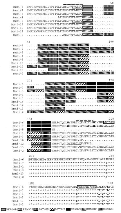
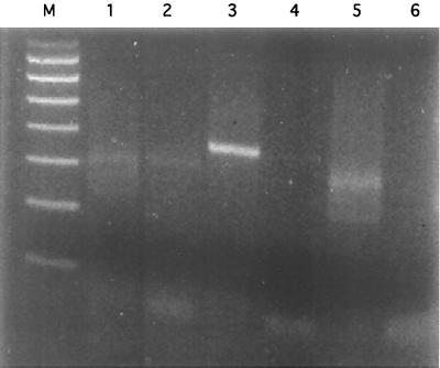
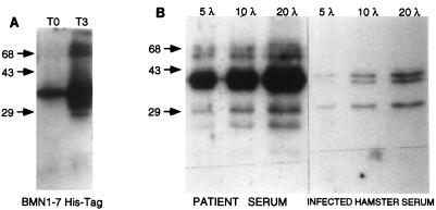
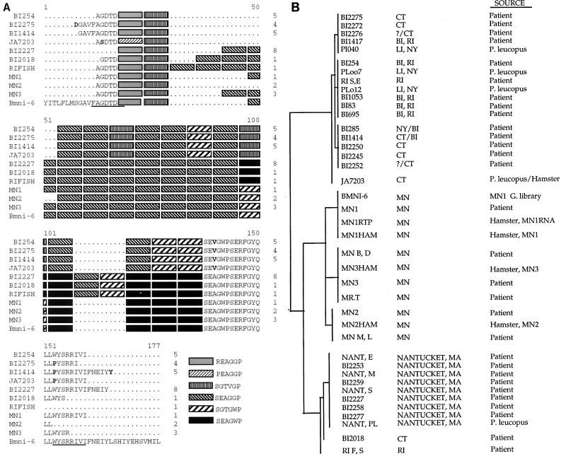
Human babesiosis in the United States is caused predominantly by Babesia microti, a tick-transmitted blood parasite. Improved testing methods for the detection of infection with this parasite are needed, since asymptomatic B. microti infection represents a potential threat to the blood supply in areas where B. microti is endemic. We performed immunoscreening of an expression library of genomic DNA from a human isolate of B. microti (strain MN1). Among 17 unique immunoreactive clones, we identified 9 which represent a related family of genes with little sequence homology to other known sequences but with an architecture resembling that of several surface proteins of Plasmodium. Within this family, a tandem array of a degenerate six-amino-acid repeat (SEAGGP, SEAGWP, SGTGWP, SGTVGP) was found in various lengths between relatively well conserved segments at the N and C termini. In order to examine within-clone variation, we developed a PCR protocol for direct recovery of a specific bmn1-6 homologue directly from 30 human blood isolates, 4 corresponding hamster isolates, and 5 geographically corresponding Peromyscus leucopus (white-footed mouse) isolates. Isolates from the hamsters had the same sequences as those found in the corresponding human blood, suggesting that genetic variation of bmn1-6 does not occur during passage. However, clones from different patients were often substantially different from each other with regard to the number and location of the degenerate repeats within the bmn1-6 homologue. Moreover, we found that strains that were closely related geographically were also closely related at the sequence level; nine patients, all from Nantucket Island, Mass., harbored clones that were indistinguishable from each other but that were distinct from those found in other northeastern or upper midwestern strains. We conclude that considerable genetic and antigenic diversity exists among isolates of B. microti from the United States and that geographic clustering of subtypes may exist. The nature of the bmn1-6 gene family suggests a mechanism of antigenic variation in B. microti that may occur by recombination, differential expression, or a combination of both mechanisms.
Figures
References
-
- Borst P, Bitter W, Blundell P A, Chaves I, Cross M, Gerrits H, van Leeuwen F, McCulloch R, Taylor M, Rudenko G. Control of VSG gene expression sites in Trypanosoma brucei. Mol Biochem Parasitol. 1998;91:67–76. - PubMed
-
- Carson C A, Brandt H M, Jensen J B, Bailey C W, Allen G K. Use of random amplified polymorphic DNA analysis to compare Babesia bovis and B. bigemina isolates. Parasitol Res. 1994;80:312–315. - PubMed
-
- Coppel R L, Culvenor J G, Bianco A E, Crewther P E, Stahl H D, Brown G V, Anders R F, Kemp D J. Variable antigen associated with the surface of erythrocytes infected with mature stages of Plasmodium falciparum. Mol Biochem Parasitol. 1986;20:265–77. - PubMed
-
- Cowman A F, Bernard O, Stewart N, Kemp D J. Genes of the protozoan parasite Babesia bovis that rearrange to produce RNA species with different sequences. Cell. 1984;37:653–660. - PubMed
Publication types
MeSH terms
Substances
Grants and funding
LinkOut - more resources
Full Text Sources
Other Literature Sources
