. 2000 Jan;38(1):382-8.
doi: 10.1128/JCM.38.1.382-388.2000.

Temporal changes in outer surface proteins A and C of the lyme disease-associated spirochete, Borrelia burgdorferi, during the chain of infection in ticks and mice

Affiliations

Temporal changes in outer surface proteins A and C of the lyme disease-associated spirochete, Borrelia burgdorferi, during the chain of infection in ticks and mice

T G Schwan et al. J Clin Microbiol. 2000 Jan.

Abstract

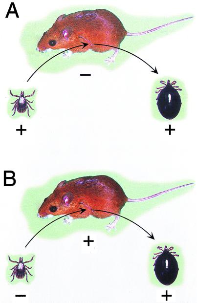
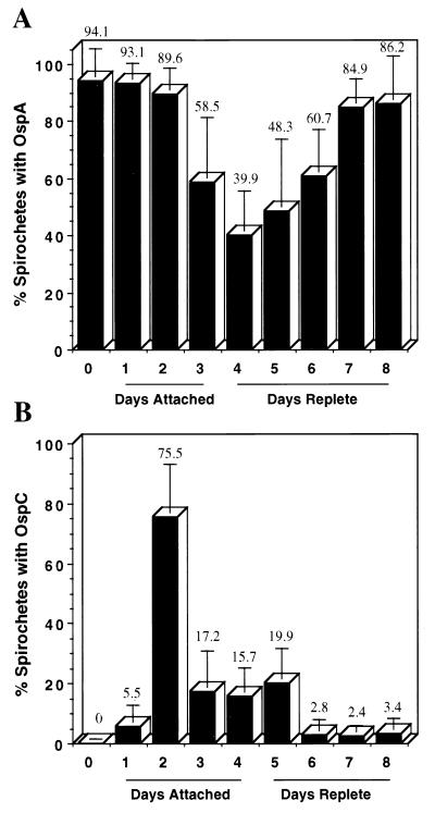
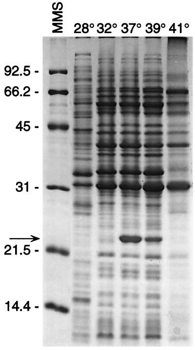
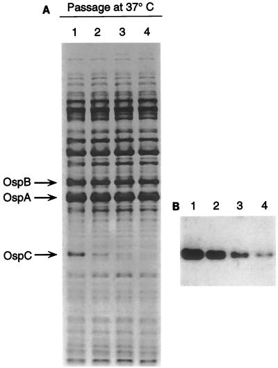
The Lyme disease-associated spirochete, Borrelia burgdorferi, is maintained in enzootic cycles involving Ixodes ticks and small mammals. Previous studies demonstrated that B. burgdorferi expresses outer surface protein A (OspA) but not OspC when residing in the midgut of unfed ticks. However, after ticks feed on blood, some spirochetes stop making OspA and express OspC. Our current work examined the timing and frequency of OspA and OspC expression by B. burgdorferi in infected Ixodes scapularis nymphs as they fed on uninfected mice and in uninfected I. scapularis larvae and nymphs as they first acquired spirochetes from infected mice. Smears of midguts from previously infected ticks were prepared at 12- or 24-h intervals following attachment through repletion at 96 h, and spirochetes were stained for immunofluorescence for detection of antibodies to OspA and OspC. As shown previously, prior to feeding spirochetes in nymphs expressed OspA but not OspC. During nymphal feeding, however, the proportion of spirochetes expressing OspA decreased, while spirochetes expressing OspC became detectable. In fact, spirochetes rapidly began to express OspC, with the greatest proportion of spirochetes having this protein at 48 h of attachment and then with the proportion decreasing significantly by the time that the ticks had completed feeding. In vitro cultivation of the spirochete at different temperatures showed OspC to be most abundant when the spirochetes were grown at 37 degrees C. Yet, the synthesis of this protein waned with continuous passage at this temperature. Immunofluorescence staining of spirochetes in smears of midguts from larvae and nymphs still attached or having completed feeding on infected mice demonstrated that OspA but not OspC was produced by these spirochetes recently acquired from mice. Therefore, the temporal synthesis of OspC by spirochetes only in feeding ticks that were infected prior to the blood meal suggests that this surface protein is involved in transmission from tick to mammal but not from mammal to tick.


Figures

FIG. 1
FIG. 1
These diagrams depict the two transmission events examined. (A) An infected nymph (+) feeding on an uninfected white-footed mouse (−). Prior to, during, and after feeding, the spirochetes in the ticks' midguts were examined for expression of OspA and OspC. (B) An uninfected nymph (−) feeding on an infected white-footed mouse (+). The spirochetes that were acquired during tick feeding and that were in the ticks' midguts were examined for expression of OspA and OspC.
FIG. 2
FIG. 2
Proportion of B. burgdorferi expressing OspA or OspC in previously infected nymphal I. scapularis ticks during attachment and feeding (as depicted in Fig. 1A). In the two separate experiments, the midguts from unfed (day 0), attached (days attached), and fully engorged (days replete) nymphal I. scapularis ticks infected prior to feeding were examined by double immunofluorescence staining to determine the percentage of spirochetes expressing OspA (A) or OspC (B). The number above each column is the mean percentage for all spirochetes examined on that sampling day. The vertical error bar represents 1 standard deviation of the mean. As shown, the spirochetes in the tick midgut displayed dramatic temporal regulation of both proteins associated with tick attachment and feeding.
FIG. 3
FIG. 3
The rapid acquisition of B. burgdorferi by nymphal I. scapularis ticks during attachment and early feeding on infected mice and the early presence of OspA (as depicted in Fig. 1B). The midguts from nymphal ticks were examined from ticks pulled off mice at 24 h (n = 11 ticks) and 48 h (n = 5 ticks) after attachment. At 24 h, 10 of 11 ticks were already infected, while at 48 h, all 5 ticks were infected. The number of spirochetes was determined by IFA with anti-OspA monoclonal antibody H5332. The mean number of spirochetes detected in all ticks at both times is shown above the column, and the vertical error bar represents 1 standard deviation of the mean.
FIG. 4
FIG. 4
Influence of different growth temperatures on the in vitro synthesis of OspC by B. burgdorferi. Spirochetes were grown in BSK-II medium to the stationary phase at each of the indicated temperatures. Whole-cell lysates of the spirochetes were examined by SDS-PAGE with proteins stained with Coomassie brilliant blue. Molecular mass standards (MMS) are shown at the left (in kilodaltons). OspC, shown to the right of the arrow, is most abundant in the culture grown at 37°C.
FIG. 5
FIG. 5
Influence of continued in vitro cultivation of B. burgdorferi at 37°C on the presence of OspC. Spirochetes were grown to the stationary phase in BSK-II medium and were passaged three consecutive times. Whole-cell lysates of the spirochetes from each passage were examined by SDS-PAGE with proteins stained with Coomassie brilliant blue (A). Immunoblot analysis with rabbit anti-OspC antiserum (B) confirmed that the protein down-regulated during passage was OspC.

References

    1. Aguero-Rosenfeld M E, Nowakowski J, Bittker S, Cooper D, Nadelman R B, Wormser G P. Evolution of the serologic response to Borrelia burgdorferi in treated patients with culture-confirmed erythema migrans. J Clin Microbiol. 1996;34:1–9. - PMC - PubMed
    1. Akins D R, Bourell K W, Caimano M J, Norgard M V, Radolf J D. A new animal model for studying Lyme disease spirochetes in a mammalian host-adapted state. J Clin Invest. 1998;101:2240–2250. - PMC - PubMed
    1. Barbour A G, Hayes S F, Heiland R A, Schrumpf M E, Tessier S L. A Borrelia-specific monoclonal antibody binds to a flagellar epitope. Infect Immun. 1986;52:549–554. - PMC - PubMed
    1. Barbour A G, Tessier S L, Todd W J. Lyme disease spirochetes and ixodid tick spirochetes share a common surface antigenic determinant defined by a monoclonal antibody. Infect Immun. 1983;41:795–804. - PMC - PubMed
    1. Barthold S W, Fikrig E, Bockenstedt L K, Persing D H. Circumvention of outer surface protein A immunity by host-adapted Borrelia burgdorferi. Infect Immun. 1995;63:2255–2261. - PMC - PubMed