. 2000 Jan 1;522 Pt 1(Pt 1):83-96.
doi: 10.1111/j.1469-7793.2000.t01-3-00083.x.

Action of a NO donor on the excitation-contraction pathway activated by noradrenaline in rat superior mesenteric artery

Affiliations

Action of a NO donor on the excitation-contraction pathway activated by noradrenaline in rat superior mesenteric artery

P Ghisdal et al. J Physiol. .

Abstract

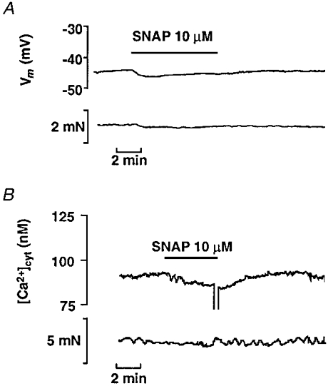
The aim of the present study was to investigate the actions of NO donors in ratsuperior mesenteric artery stimulated with noradrenaline by studying their effects on isometric tension, membrane potential (Vm), cytosolic calcium concentration ([Ca2+]cyt) and accumulation of inositol phosphates. In unstimulated arteries, SNAP (S-nitroso-N-acetylpenicillamine, 10 microM) hyperpolarised Vm by 3.0 +/- 0.5 mV (n = 9). In KCl-stimulated arteries, SNAP relaxed contraction without changing Vm and [Ca2+]cyt. In noradrenaline-stimulated arteries, SNAP relaxed tension, repolarised Vm and decreased [Ca2+]cyt with the same potency. Responses to SNAP were unaffected by the following K+ channel blockers: glibenclamide, 4-aminopyridine, apamin and charybdotoxin, and by increasing the KCl concentration to 25 mM. In SNAP-pretreated arteries, the production of inositol phosphates and the contraction stimulated by noradrenaline were inhibited similarly. The guanylate cyclase inhibitor ODQ abolished the increase in cyclic GMP content evoked by SNAP and inhibited the effects of SNAP on contraction, Vm and accumulation of inositol phosphates in noradrenaline-stimulated artery. These results indicate that, in rat superior mesenteric arteries activated by noradrenaline, inhibition of production of inositol phosphates is responsible for the effects of the NO donor SNAP on membrane potential, [Ca2+]cyt and contraction through a cyclic GMP-dependent mechanism.


Figures

Figure 1
Figure 1. Effects of SNAP in unstimulated mesenteric artery
A, representative experimental traces showing simultaneous records of smooth muscle membrane potential (Vm, upper trace) and tension (lower trace) in an unstimulated artery segment. SNAP was applied as indicated. B, representative experimental traces showing simultaneous records of cytosolic calcium concentration ([Ca2+]cyt, upper trace) and tension (lower trace) in an unstimulated artery segment loaded with fura-2.
Figure 2
Figure 2. Effects of SNAP on membrane potential, cytosolic Ca2+ concentration and contractile tension in KCl-stimulated mesenteric artery
A, left panel: representative experimental traces of simultaneous records of membrane potential (Vm, upper trace) and tension (lower trace) showing the lack of repolarisation and the marked relaxation evoked by SNAP (10 μm) in a mesenteric artery segment depolarised and contracted by 40 mM KCl. Right panel: mean values +s.e.m. from 6 arteries. Responses to KCl were measured immediately before the addition of SNAP. Responses to SNAP were measured at their maximum. * P < 0.05 versus KCl. B, left panel: representative traces showing simultaneous records of cytosolic Ca2+ concentration ([Ca2+]cyt, upper trace) and contractile tension (lower trace), in a mesenteric artery segment stimulated with KCl (40 mM). SNAP (10 μm) was applied as indicated. Right panel: mean values +s.e.m. from 5 arteries. Responses to KCl were measured immediately before the addition of SNAP. Responses to SNAP were measured at their maximum. * P < 0.05 versus KCl.
Figure 3
Figure 3. Effects of SNAP on membrane potential, cytosolic Ca2+ concentration and contractile tension in noradrenaline-stimulated mesenteric artery
A, representative original traces of simultaneous records of membrane potential (Vm, upper trace) and contractile tension (lower trace) showing the repolarisation and the relaxation induced by SNAP (10 μm) in an artery segment depolarised and contracted by noradrenaline (NA, 0.5 μm). B, experimental traces showing simultaneous records of cytosolic calcium concentration ([Ca2+]cyt, upper trace) and contractile tension (lower trace) in a fura-2-loaded mesenteric artery segment stimulated with noradrenaline (NA, 1 μm) C, dose-effect curves for the effects of SNAP on membrane potential and tension. The change in membrane potential (expressed in mV) is compared with the relaxation (expressed as a percentage of the contraction to noradrenaline) evoked by increasing concentrations of SNAP applied on the plateau of the contraction in untreated arteries (□, change in membrane potential, ΔVm; ○, relaxation) and in arteries preincubated in the presence of the guanylate cyclase inhibitor ODQ (10 μm) (▪, change in membrane potential, ΔVm; •, relaxation). The dashed line represents the change in potential reversing the depolarisation evoked by noradrenaline. Data are mean values ±s.e.m. from 7 experiments. D, dose-effect curves for the effects of SNAP on cytosolic Ca2+ concentration (▵) and tension (○). Increasing concentrations of SNAP were cumulatively applied on arteries stimulated with noradrenaline (0.5-1 μm). The change in cytosolic Ca2+ concentration and the relaxation are expressed as a percentage of the response to noradrenaline measured before the addition of SNAP. Data are mean values ±s.e.m. from 6 experiments.
Figure 4
Figure 4. Effects of K+ channel blockers on the repolarisation and the relaxation in response to SNAP (10 μm) in arteries contracted with noradrenaline
A-D, left panels: original records showing the changes in membrane potential (Vm, upper trace) and in tension (lower trace) induced by SNAP in the presence of charybdotoxin (Ctx, 0.1 μm) and apamin (Apa, 0.1 μm; A), 4-aminopyridine (4-AP, 0.5 mm; B), glibenclamide (Gli, 0.1 μm; C), and a cocktail of charybdotoxin (0.1 μm) + apamin (0.1 μm) + 4-aminopyridine (5 mM) + glibenclamine (10 μm; D), in artery segments stimulated by noradrenaline (NA, 0.5 μm). Right panels: bar graphs showing the mean values +s.e.m. of the change in membrane potential (Vm) evoked by the blocker(s) (□), noradrenaline in the presence of the blocker(s) before the addition of SNAP (▪) and after the addition of SNAP (1–10 μm) (formula image,formula image). The base of the columns corresponds to the level of the resting membrane potential. Data are means +s.e.m. from 4–11 recordings.
Figure 5
Figure 5. Effects of 25 mM KCl on the change in membrane potential and in cytosolic Ca2+ concentration and on the relaxation evoked by SNAP in noradrenaline-stimulated mesenteric artery
A, left panel: original records showing the changes in membrane potential (Vm, upper trace) and in tension (lower trace) induced by SNAP in a mesenteric artery ring stimulated with noradrenaline in the presence of 25 mm KCl. Right panel: mean values +s.e.m. from 4 recordings of the membrane potential (Vm). The base of the columns corresponds to the level of the resting membrane potential. B, left panel: original traces showing simultaneous records of cytosolic Ca2+ concentration ([Ca2+]cyt, upper trace) and contractile tension (lower trace), in a mesenteric artery segment stimulated with noradrenaline in the presence of 25 mM KCl. SNAP (10 μm) and phentolamine (Phent, 10 μm) were applied as indicated. Right panel: mean values +s.e.m. from 3 recordings of the change in [Ca2+]cyt.
Figure 6
Figure 6. Effect of SNAP on inositol phosphates accumulation in noradrenaline-stimulated preparation
A,[3H]myo-inositol-loaded arteries were pretreated with SNAP and/or ODQ (10 μm) for 1 min and further incubated in the presence of noradrenaline for 30 min. Data are from a typical experiment, performed in triplicate. * P < 0.05 versus NA in the absence of SNAP. B, comparison of the inhibition by SNAP of inositol phosphates accumulation and of contraction evoked by noradrenaline. Arteries were pretreated with SNAP for 1 min before the addition of noradrenaline. Contraction was measured by the area under the curve. Data are means ±s.e.m. from 6 determinations.
Figure 7
Figure 7. Effects of SNAP on membrane potential, cytosolic Ca2+ concentration and contractile tension in mesenteric artery segments stimulated with AlF4
A, left panel: experimental traces of simultaneous records of membrane potential (Vm, upper trace) and tension (lower trace) showing the repolarisation and the relaxation induced by SNAP (10 μm) in an artery segment depolarised and contracted with AlF4. Right panel: mean values +s.e.m. from 4 recordings. Responses to AlF4 were measured immediately before the addition of SNAP. Responses to SNAP were measured at their maximum. ** P < 0.01. B, left panel: experimental traces showing simultaneous records of cytosolic calcium concentration ([Ca2+]cyt, upper trace) and contractile tension (lower trace) in a fura-2-loaded artery segment stimulated with AlF4. Right panel: mean values +s.e.m. from 4 recordings. Responses to AlF4 were measured immediately before the addition of SNAP. Responses to SNAP were measured at their maximum. ** P < 0.01.
Figure 8
Figure 8. Effects of cromakalim on the depolarisation and the contraction evoked by noradrenaline
Increasing concentrations of cromakalim were cumulatively applied on arteries stimulated with noradrenaline (0.5 μm). Membrane potential (□) and contractile tension (○) were recorded simultaneously. Changes in membrane potential are expressed as the difference between the membrane potential value recorded in the presence of cromakalim and its value recorded in the presence of noradrenaline before the addition of cromakalim. The relaxation is expressed as a percentage of the contraction evoked by noradrenaline. The dashed line represents the change in potential reversing completely the depolarisation evoked by noradrenaline. Data are mean values ±s.e.m. from 6 experiments.

Similar articles

Cited by

References

    1. Alosachie I, Godfraind T. The modulatory role of vascular endothelium in the interaction of agonists and antagonists with α-adrenoceptors in the rat aorta. British Journal of Pharmacology. 1988;95:619–629. - PMC - PubMed
    1. Amédée T, Benham CD, Bolton TB, Byrne NG, Large WA. Potassium, chloride and non-selective cation conductances opened by noradrenaline in rabbit ear artery cells. The Journal of Physiology. 1990;423:551–568. - PMC - PubMed
    1. Archer SL, Huang JMC, Hampl V, Nelson DP, Schultz PJ, Weir EK. Nitric oxide and cGMP cause vasorelaxation by activation of a charybdotoxin-sensitive K channel by cGMP-dependent protein kinase. Proceedings of the National Academy of Sciences of the USA. 1994;91:7583–7587. - PMC - PubMed
    1. Archer SL, Huang JMC, Reeve HL, Hampl V, Tolarova S, Michelakis E, Weir EK. Differential distribution of electrophysiologically distinct myocytes in conduit and resistance arteries determines their response to nitric oxide and hypoxia. Circulation Research. 1996;78:431–442. - PubMed
    1. Armstead WM. Role of activation of calcium sensitive K+ channels in NO and hypoxia induced pial artery vasodilation. American Journal of Physiology. 1997;272:H1785–1790. - PubMed

Publication types

MeSH terms

LinkOut - more resources