. 2000 Jan;66(1):23-8.
doi: 10.1128/AEM.66.1.23-28.2000.

Purification and partial characterization of a murein hydrolase, millericin B, produced by Streptococcus milleri NMSCC 061

Affiliations

Purification and partial characterization of a murein hydrolase, millericin B, produced by Streptococcus milleri NMSCC 061

M Beukes et al. Appl Environ Microbiol. 2000 Jan.

Abstract

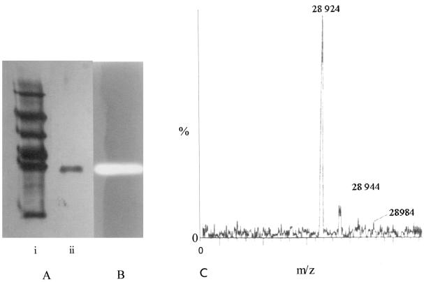
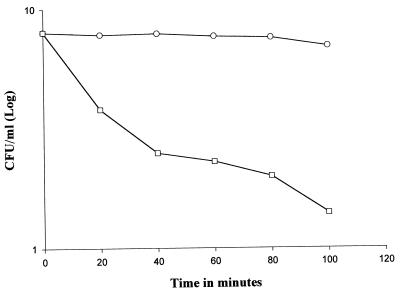
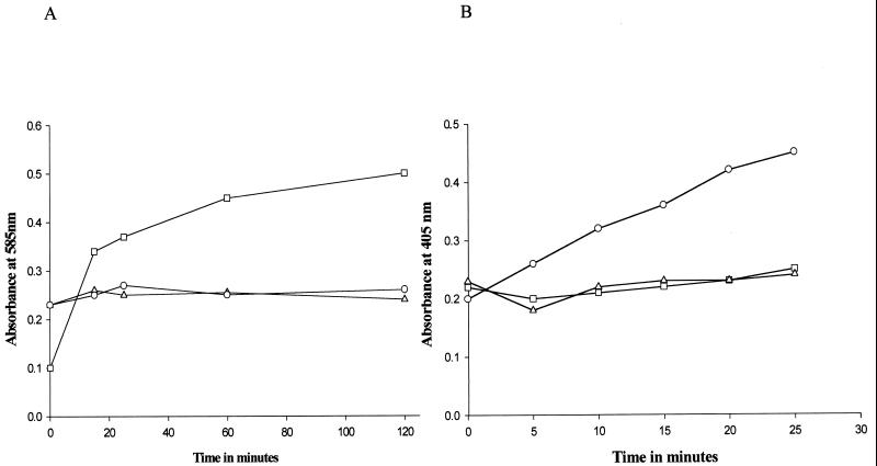
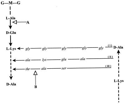
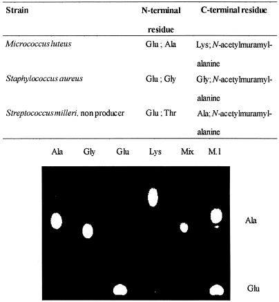
Streptococcus milleri NMSCC 061 was screened for antimicrobial substances and shown to produce a bacteriolytic cell wall hydrolase, termed millericin B. The enzyme was purified to homogeneity by a four-step purification procedure that consisted of ammonium sulfate precipitation followed by gel filtration, ultrafiltration, and ion-exchange chromatography. The yield following ion-exchange chromatography was 6.4%, with a greater-than-2,000-fold increase in specific activity. The molecular weight of the enzyme was 28,924 as determined by electrospray mass spectrometry. The amino acid sequences of both the N terminus of the enzyme (NH(2) SENDFSLAMVSN) and an internal fragment which was generated by cyanogen bromide cleavage (NH(2) SIQTNAPWGL) were determined by automated Edman degradation. Millericin B displayed a broad spectrum of activity against gram-positive bacteria but was not active against Bacillus subtilis W23 or Escherichia coli ATCC 486 or against the producer strain itself. N-Dinitrophenyl derivatization and hydrazine hydrolysis of free amino and free carboxyl groups liberated from peptidoglycan digested with millericin B followed by thin-layer chromatography showed millericin B to be an endopeptidase with multiple activities. It cleaves the stem peptide at the N terminus of glutamic acid as well as the N terminus of the last residue in the interpeptide cross-link of susceptible strains.


Figures

FIG. 1
FIG. 1
(A) Polyacrylamide (12%) gel electrophoresis of purified millericin. Lanes: i, mid-range molecular weight standards (Promega); ii, purified millericin after ion-exchange chromatography. (B) Gel slice of a 12% acrylamide zymogram containing 0.1% M. luteus cell walls stained with 0.1% methylene blue in 0.01% KOH following renaturation of millericin PB in 0.1% Triton X-100 and 10 mM MgCl2. (C) Electrospray mass spectrometry of millericin B after ion-exchange chromatography.
FIG. 2
FIG. 2
Effect of millericin B on the growth of M. luteus ATCC 46898. Symbols: ○, control (addition of MQ water); □, test (addition of millericin).
FIG. 3
FIG. 3
(A) Liberation of free reducing sugars from the cell walls of M. luteus ATCC 46898 after digestion with a cell wall-degrading enzyme, analyzed by the Morgan-Elson test (12). Symbols: □, cell walls digested with lysozyme; ○, cell walls digested with millericin B; ▵, cell wall in the presence of 20 mM phosphate buffer (pH 7.0). (B). Liberation of free amino groups from the digestion of M. luteus ATCC 46898 cell walls with millericin B by N-dinitrophenol (DNP) derivatization (12). Symbols: ○, cell walls digested with millericin B; □, cell walls digested with lysozyme; ▵, cell walls in the presence of 20 mM phosphate buffer (pH 7.0).
FIG. 4
FIG. 4
Fragments of the primary structure, stem peptide, and interpeptide cross bridge of peptidoglycans found in S. aureus (i), M. luteus (ii), and S. milleri (iii). A and B, cleavage sites of millericin B as determined by DNP derivatization and DNP-hydrazine derivatization.
FIG. 5
FIG. 5
Amino acid residues detected after digestion of peptidoglycan from three bacterial strains, DNP derivatization was used to detect free N termini and DNP-hydrazine derivatization was used for the C termini. A representative thin-layer chromatograph of cell walls from M. luteus appears as an example. Mix, mixture of standard amino acids that occur in the cell wall of M. luteus; M.l., M. luteus.

References

    1. Amako K, Umeda A, Murata K. Arrangement of peptidoglycan in the cell wall of Staphylococcus spp. J Bacteriol. 1982;150:844–850. - PMC - PubMed
    1. Araki Y, Nakatani T, Wakayama K, Ito E. Occurrence of N-nonsubstituted glucosamine residues in peptidoglycan of lysozyme resistant cell walls of Bacillus cereus. J Biol Chem. 1972;247:6312–6322. - PubMed
    1. Bierbaum G, Sahl H-G. Autolytic system of Staphylococcus simulans 22: influence of cationic peptides on activity of N-acetylmuramoyl-l-alanine amidase. J Bacteriol. 1987;169:5452–5458. - PMC - PubMed
    1. Browder H P, Zygmunt W A, Young J R, Tavorina P A. Lysostaphin: enzymatic mode of action. Biochem Biophys Res Commun. 1965;19:383–389. - PubMed
    1. Clarke A J. Extent of peptidoglycan O-acetylation in the tribe Proteae. J Bacteriol. 1993;175:4550–4553. - PMC - PubMed

Publication types

MeSH terms

Substances

LinkOut - more resources