. 2000 Jan;83(1):69-75.
doi: 10.1136/heart.83.1.69.

Role of endothelial cell denudation and smooth muscle cell dedifferentiation in neointimal formation of human vein grafts after coronary artery bypass grafting: therapeutic implications

Affiliations

Role of endothelial cell denudation and smooth muscle cell dedifferentiation in neointimal formation of human vein grafts after coronary artery bypass grafting: therapeutic implications

Y Sasaki et al. Heart. 2000 Jan.

Abstract

Objective: To provide better insights into the genesis of neointimal thickening in human vein grafts early after surgery.

Design: Retrospective study.

Setting: Tertiary referral centre.

Subjects: 18 distal anastomotic sites of patent grafts, obtained at necropsy from eight patients who died over differing periods (ranging from two days to nine months) after the procedure.

Main outcome measures: Immunohistochemical evaluation of smooth muscle cell phenotype modulation in relation to proliferative activity.

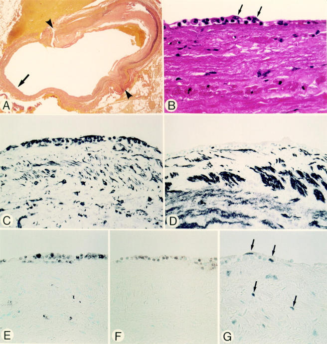
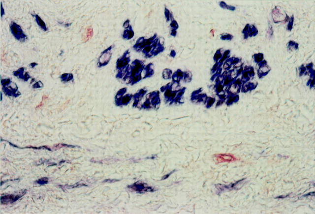
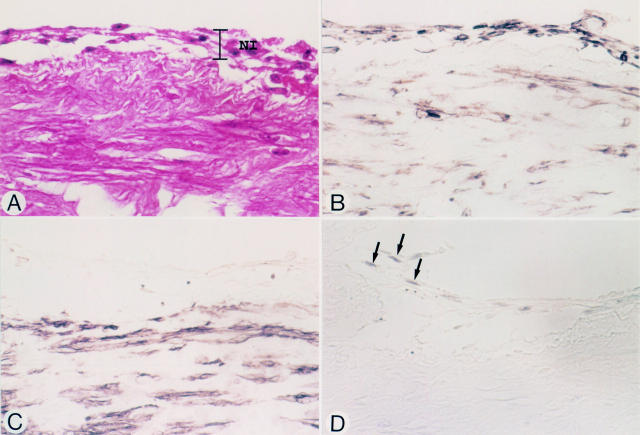
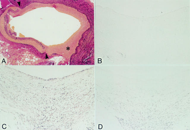
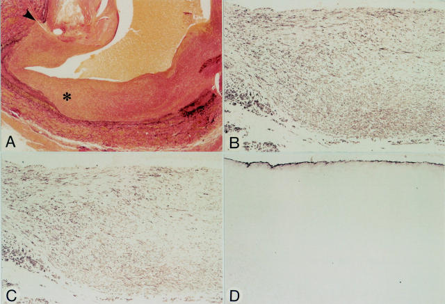
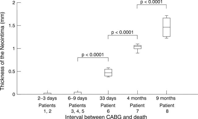
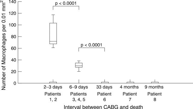
Results: The earliest changes are characterised by loss of surface lining endothelial cells and insudation of blood corpuscular elements admixed with fibrin-platelet thrombus. At sites of injury vimentin positive and actin negative spindle shaped cells appear in the intima, while the related pre-existent media shows focal absence of actin positive smooth muscle cells. Proliferative activity colocalises at these sites. With time distinct neointimal thickening occurs, associated with disappearance of proliferative activity and a phenotypic shift of the smooth muscle cells.

Conclusions: The observation that luminal endothelial cell denudation, with insudation of the intima with blood elements, occurs in the very early stages suggests that these phenomena are responsible for the observed dedifferentiation of pre-existent smooth muscle cells, known to be a prerequisite for cell proliferation and the evolution of intimal thickening. It is likely, therefore, that platelet released growth factors play a pivotal role, which thus may provide a target for preventive pharmacological intervention.


Figures

Figure 1
Figure 1
Micrographs of an anastomotic site, taken two days after grafting (patient 1). Panels A-G are serial sections. (A) Elastic tissue stain. The site of anastomosis, indicated by arrowheads, with the right coronary artery. The luminal surface of the vein graft shows a cellular response. Details of the cellular response, indicated by the arrow, are shown in panels B-G. (B) Haematoxylin and eosin stain. At the luminal surface endothelial cells have been denuded. The earliest cellular response of the grafts is demonstrated by an accumulation of polymorphonuclear leucocytes and mononuclear round cells, amid a fibrin-platelet thrombus, partially covered by spindle shaped cells (arrows). (C) Vimentin stain. Spindle shaped cells and round cells at the response site are positive. (D) HHF-35. The spindle shaped cells and round cells are negative. (E) HAM-56. Some round cells stain with this macrophage marker. None of the spindle shaped cells at the luminal site stain positive. (F) UCHL-1. Some small round cells stain with this T lymphocyte marker. (G) PCNA. Positive cells are seen at the site of cellular response and in the adjacent pre-existent media. Original magnification: (A) × 18; (B) × 580; (C-G) × 360.
Figure 2
Figure 2
Double immunostaining with HHF-35 (blue) and PCNA (red) of an anastomotic site, two days after grafting (patient 1). The media contains a few PCNA positive cells (red), which lack staining for actin and most likely represent dedifferentiated SMCs. Most SMCs are actin positive (blue). Original magnification × 720.
Figure 3
Figure 3
Micrographs of a vein graft at an anastomotic site, nine days after grafting (patient 5). Panels A-D represent serial sections. (A) Haematoxylin and eosin stain. Endothelial cells have been denuded. Early neointimal tissue (NI) is seen at the luminal surface of the graft. (B) Vimentin stain. Both spindle shaped cells and round cells in the neointima are positive. (C) HHF-35. The cells in the neointima do not stain. (D) PCNA. Some of the HHF-35 negative spindle shaped cells (arrows) stain positive. Original magnification × 580.
Figure 4
Figure 4
Micrographs of an anastomotic site, 33 days after grafting (patient 6). Panels A-D represent serial sections. (A) Elastic tissue stain. The anastomotic site (arrowheads) to the obtuse marginal branch shows distinct neointimal proliferation of the vein graft (asterisk). (B) HAM-56. No positivity for macrophages in the neointima. (C) HHF-35. Spindle shaped cells in the neointima stain positive. (D) CGA-7. Spindle shaped cells within the deeper layers of the neointima stain positive, but those closer to the luminal site are negative suggesting that these cells have not yet fully differentiated. The anti-vWf antibody was negative at the luminal side (not shown). Original magnification: (A) × 36; (B-D) × 145.
Figure 5
Figure 5
Micrographs of an anastomotic site, nine months after grafting (patient 8). Panels A-D represent serial sections. (A) Elastic tissue stain. Note distinct neointimal tissue at the anastomotic site (arrowhead). The neointima at the site of the asterisk is shown at higher magnification in panels B-D. (B) HHF-35. Spindle shaped cells in the neointima are positive. (C) CGA-7. Most spindle shaped cells are positive suggesting that most cells have the phenotype of fully differentiated SMCs (compare to panel B). (D) Anti-vWf. Regenerated endothelial cells line the luminal surface. Original magnification: (A) × 30; (B-D) × 90.
Figure 6
Figure 6
Thickness of the neointima.
Figure 7
Figure 7
Number of macrophages within the neointima.

References

    1. J Cell Biol. 1985 Mar;100(3):807-13 - PubMed
    1. Arterioscler Thromb Vasc Biol. 1997 Dec;17(12):3611-9 - PubMed
    1. Am Heart J. 1990 May;119(5):1164-84 - PubMed
    1. Circulation. 1991 Apr;83(4):1327-32 - PubMed
    1. Circulation. 1971 Oct;44(4):719-28 - PubMed

Substances