. 2000 Jan 4;97(1):85-90.
doi: 10.1073/pnas.97.1.85.

The crystal structure of modified bovine fibrinogen

Affiliations

The crystal structure of modified bovine fibrinogen

J H Brown et al. Proc Natl Acad Sci U S A. .

Abstract

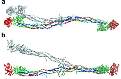
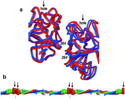
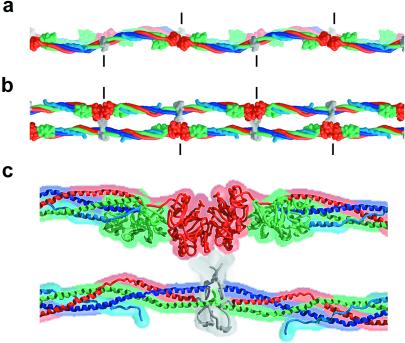
Here we report the crystal structure at approximately 4-A resolution of a selectively proteolyzed bovine fibrinogen. This key component in hemostasis is an elongated 340-kDa glycoprotein in the plasma that upon activation by thrombin self-assembles to form the fibrin clot. The crystals are unusual because they are made up of end-to-end bonded molecules that form flexible filaments. We have visualized the entire coiled-coil region of the molecule, which has a planar sigmoidal shape. The primary polymerization receptor pockets at the ends of the molecule face the same way throughout the end-to-end bonded filaments, and based on this conformation, we have developed an improved model of the two-stranded protofibril that is the basic building block in fibrin. Near the middle of the coiled-coil region, the plasmin-sensitive segment is a hinge about which the molecule adopts different conformations. This segment also includes the boundary between the three- and four-stranded portions of the coiled coil, indicating the location on the backbone that anchors the extended flexible Aalpha arm. We suggest that a flexible branch point in the molecule may help accommodate variability in the structure of the fibrin clot.


Figures

Figure 1
Figure 1
Three perpendicular views of the modified bovine fibrinogen molecule. Color scheme (for all figures) is blue for the Aα-chain, green for the Bβ-chain, and red for the γ-chain. The antiparallel portion of the Aα-chain is shown in light blue. The chains in the central N-terminal disulfide knot region cannot be traced and their general location is indicated in gray. Carbohydrates are indicated by CH2O. (Bovine residue numbers are indicated, see Methods.) (a) View showing the sigmoidal shape of the coiled-coil axis. (b) View perpendicular to that in a indicating the molecular planarity. Arrow indicates approximate 2-fold axis of the molecule, which is nearly perpendicular to the plane of the sigmoidal coiled coil. Observed C termini are indicated in italics. (c and d) Cross-sectional views of the four-stranded coiled coil at locations indicated in b. The four-stranded coiled coil is quite asymmetric and may be better described as an overlapping pair of three-stranded coiled coils that share the parallel Aα- and Bβ-chains. (The γ and antiparallel Aα-chains do not contact one another.) (e) Magnified view of b showing part of the coiled coil superimposed on an electron density map (see Methods). A polyalanine model in this segment of the antiparallel Aα-chain density (Bottom, light blue) is shown. Regular helical density is observed for the parallel chains.
Figure 2
Figure 2
Conformational flexibility of fibrinogen in the crystals. The diagrams show superpositions of noncrystallographically related fibrinogen molecules based on the least-squares fit of the relatively rigid coiled-coil segment: Aα104-Aα154, Bβ140-Bβ190, γ77-γ127. Among the different noncrystallographically related copies, the rms difference between the coordinates of this segment is about half that of the backbone's most flexible segment: Aα64-Aα114, Bβ100-Bβ150, γ37-γ87. (a) View, as in Fig. 1a, of one pair of molecules whose conformations differ primarily by bending within the plane of the sigmoidal coiled-coil axis. (b) View, as in Fig. 1b, of a different pair of molecules whose conformations differ primarily by bending out of the plane of the sigmoidal axis.
Figure 3
Figure 3
Conserved end-to-end molecular interactions. (a) Superposition of six γ-domain dimers derived from the various crystals of modified bovine fibrinogen (red) and human fragment D and crosslinked D-dimer (blue) (24, 25) show the γ domains to be similarly “offset” from one another. This feature can be visualized by noting, for example, that γ264 of the right monomer is interacting at the edge of the γ-γ interface whereas in the left monomer it is interacting at the center of the interface. No significant difference in the offset is found among the three bovine γ-domain dimers or among the three human γ-domain dimers (pooled intra-species SD is 0.455 Å). Interspecies amino acid differences at or near the interface (e.g., γ264, which is methionine in human and serine in bovine fibrinogen) probably perturb the docking of the domains, creating a slightly less staggered offset (≈1.7-Å rms difference) in the bovine γ-dimer relative to that in the human dimer. (b) Crystal structure of an end-to-end bonded fibrinogen filament. All γ-domain receptor pockets (shown by arrows) are on the same face of the extended filament.
Figure 4
Figure 4
A model of the two-stranded protofibril of fibrin derived from a filament in the bovine fibrinogen crystal structure. (a) View as in Fig. 1a. The sigmoidal coiled-coil axes of the two filaments are in phase. (b) View as in Fig. 1b. (c) Magnified view of b, including the DDE cluster. The model was constructed so that the two filaments are half-staggered (by 225 Å), closed (the γ-domain receptor pockets face the adjacent filament), and separated by ≈60 Å (a rough estimate from figure 9 in ref. and J. Weisel, personal communication). The contact area between the D and E regions is not known and is illustrated schematically here. Short vertical lines indicate the molecular boundaries. The ≈30-Å distance between the N terminus of the Aα-chain coiled coil from one filament and the γ-domain hole on the other filament (into which the GPR sequence binds) could easily be spanned by the 30 additional residues in the disulfide knot region. Similarly, the ≈100-Å distance between the N terminus of the Bβ-chain coiled coil and the β-domain receptor pocket of the adjacent filament also could be bridged by the 64 residues in this region. This long distance between the disulfide knot and the GHR sequence of the Bβ-chain also may allow binding between protofibrils.

References

    1. Doolittle R F, Everse S J, Spraggon G. FASEB J. 1996;10:1464–1470. - PubMed
    1. Hall C E, Slayter H S. J Biophys Biochem Cytol. 1959;5:11–17. - PMC - PubMed
    1. Mosesson M W, Hainfeld J, Wall J, Haschemeyer R H. J Mol Biol. 1981;153:695–718. - PubMed
    1. Erickson H P, Fowler W E. In: Molecular Biology of Fibrinogen and Fibrin. Mosesson M W, Doolittle R F, editors. New York: N.Y. Acad. Sci.; 1983. pp. 146–163.
    1. Williams R C. In: Molecular Biology of Fibrinogen and Fibrin. Mosesson M W, Doolittle R F, editors. New York: N.Y. Acad. Sci.; 1983. pp. 180–193.

Publication types

Associated data