

Substitutions in the aspartate transcarbamoylase domain of hamster CAD disrupt oligomeric structure
- PMID: 10618377
- PMCID: PMC26622
- DOI: 10.1073/pnas.97.1.97
Substitutions in the aspartate transcarbamoylase domain of hamster CAD disrupt oligomeric structure
Abstract
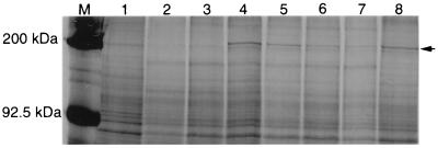
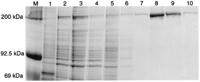
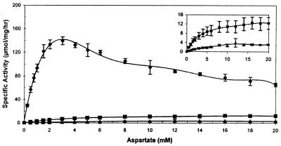
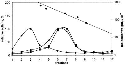
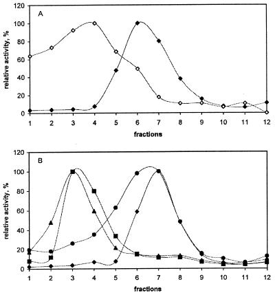
Aspartate transcarbamoylase (ATCase; EC 2.1.3.2) is one of three enzymatic domains of CAD, a protein whose native structure is usually a hexamer of identical subunits. Alanine substitutions for the ATCase residues Asp-90 and Arg-269 were generated in a bicistronic vector that encodes a 6-histidine-tagged hamster CAD. Stably transfected mammalian cells expressing high levels of CAD were easily isolated and CAD purification was simplified over previous procedures. The substitutions reduce the ATCase V(max) of the altered CADs by 11-fold and 46-fold, respectively, as well as affect the enzyme's affinity for aspartate. At 25 mM Mg(2+), these substitutions cause the oligomeric CAD to dissociate into monomers. Under the same dissociating conditions, incubating the altered CAD with the ATCase substrate carbamoyl phosphate or the bisubstrate analogue N-phosphonacetyl-L-aspartate unexpectedly leads to the reformation of hexamers. Incubation with the other ATCase substrate, aspartate, has no effect. These results demonstrate that the ATCase domain is central to hexamer formation in CAD and suggest that the ATCase reaction mechanism is ordered in the same manner as the Escherichia coli ATCase. Finally, the data indicate that the binding of carbamoyl phosphate induces conformational changes that enhance the interaction of CAD subunits.
Figures
Similar articles
-
Expression, purification, crystallization and preliminary X-ray diffraction analysis of the aspartate transcarbamoylase domain of human CAD.Acta Crystallogr Sect F Struct Biol Cryst Commun. 2013 Dec;69(Pt 12):1425-30. doi: 10.1107/S1744309113031114. Epub 2013 Nov 29. Acta Crystallogr Sect F Struct Biol Cryst Commun. 2013. PMID: 24316846 Free PMC article.
-
Half of Saccharomyces cerevisiae carbamoyl phosphate synthetase produces and channels carbamoyl phosphate to the fused aspartate transcarbamoylase domain.J Biol Chem. 1999 Aug 20;274(34):23794-801. doi: 10.1074/jbc.274.34.23794. J Biol Chem. 1999. PMID: 10446140
-
Proteolytic cleavage of the multienzyme polypeptide CAD to release the mammalian aspartate transcarbamoylase. Biochemical comparison with the homologous Escherichia coli catalytic subunit.Eur J Biochem. 1994 Nov 1;225(3):845-53. doi: 10.1111/j.1432-1033.1994.0845b.x. Eur J Biochem. 1994. PMID: 7957221
-
CAD gene sequence and the domain structure of the mammalian multifunctional protein CAD.Biochem Soc Trans. 1993 Feb;21(1):186-91. doi: 10.1042/bst0210186. Biochem Soc Trans. 1993. PMID: 8095469 Review. No abstract available.
-
New mechanisms of gene amplification in drug resistance (the episome model).Cancer Treat Res. 1991;57:1-11. doi: 10.1007/978-1-4615-3872-1_1. Cancer Treat Res. 1991. PMID: 1686710 Review. No abstract available.
Cited by
-
The human Rad9 checkpoint protein stimulates the carbamoyl phosphate synthetase activity of the multifunctional protein CAD.Nucleic Acids Res. 2004 Aug 23;32(15):4524-30. doi: 10.1093/nar/gkh789. Print 2004. Nucleic Acids Res. 2004. PMID: 15326225 Free PMC article.
-
Epileptic encephalopathy in a young Bengal cat caused by CAD deficiency.Sci Rep. 2025 Apr 18;15(1):13506. doi: 10.1038/s41598-025-98414-0. Sci Rep. 2025. PMID: 40251393 Free PMC article.
-
Deciphering CAD: Structure and function of a mega-enzymatic pyrimidine factory in health and disease.Protein Sci. 2021 Oct;30(10):1995-2008. doi: 10.1002/pro.4158. Epub 2021 Jul 22. Protein Sci. 2021. PMID: 34288185 Free PMC article. Review.
-
A Tailored Strategy to Crosslink the Aspartate Transcarbamoylase Domain of the Multienzymatic Protein CAD.Molecules. 2023 Jan 9;28(2):660. doi: 10.3390/molecules28020660. Molecules. 2023. PMID: 36677714 Free PMC article.
-
Allosteric regulation of CAD modulates de novo pyrimidine synthesis during the cell cycle.Nat Metab. 2023 Feb;5(2):277-293. doi: 10.1038/s42255-023-00735-9. Epub 2023 Feb 6. Nat Metab. 2023. PMID: 36747088 Free PMC article.
References
-
- Coleman P F, Suttle D P, Stark G R. J Biol Chem. 1977;252:6379–6385. - PubMed
-
- Evans D R. In: Multidomain Proteins: Structure and Evolution. Hardie D G, Coggins J R, editors. New York: Elsevier; 1986. pp. 283–331.
-
- Stevens R C, Chook Y M, Cho C Y, Lipscomb W N, Kantrowitz E R. Protein Eng. 1991;4:391–408. - PubMed
-
- Grayson D R, Evans D R. J Biol Chem. 1983;258:4123–4129. - PubMed
Publication types
MeSH terms
Substances
Grants and funding
LinkOut - more resources
Full Text Sources
Molecular Biology Databases
Miscellaneous
