. 2000 Jan 4;97(1):228-33.
doi: 10.1073/pnas.97.1.228.

Diverse karyotypic abnormalities of the c-myc locus associated with c-myc dysregulation and tumor progression in multiple myeloma

Affiliations

Diverse karyotypic abnormalities of the c-myc locus associated with c-myc dysregulation and tumor progression in multiple myeloma

Y Shou et al. Proc Natl Acad Sci U S A. .

Abstract

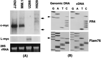
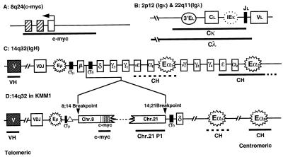
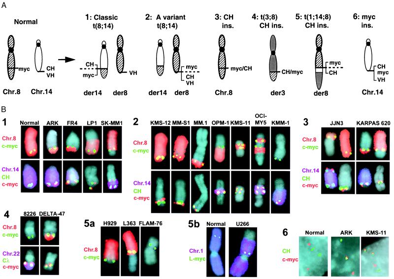
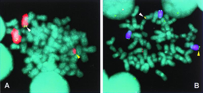
Translocations involving c-myc and an Ig locus have been reported rarely in human multiple myeloma (MM). Using specific fluorescence in situ hybridization probes, we show complex karyotypic abnormalities of the c-myc or L-myc locus in 19 of 20 MM cell lines and approximately 50% of advanced primary MM tumors. These abnormalities include unusual and complex translocations and insertions that often juxtapose myc with an IgH or IgL locus. For two advanced primary MM tumors, some tumor cells contain a karyotypic abnormality of the c-myc locus, whereas other tumor cells do not, indicating that this karyotypic abnormality of c-myc occurs as a late event. All informative MM cell lines show monoallelic expression of c-myc. For Burkitt's lymphoma and mouse plasmacytoma tumors, balanced translocation that juxtaposes c-myc with one of the Ig loci is an early, invariant event that is mediated by B cell-specific DNA modification mechanisms. By contrast, for MM, dysregulation of c-myc apparently is caused principally by complex genomic rearrangements that occur during late stages of MM progression and do not involve B cell-specific DNA modification mechanisms.


Figures

Figure 1
Figure 1
Expression of c-myc and L-myc in MM cell lines. (A) A Northern blot containing 5 μg of total RNA from four HMCL was probed sequentially with c-myc (Top) and L-myc (Middle). The normal 2.4-kb c-myc mRNA is seen in JJN3 and MM-S1, and the normal 3.6-kb L-myc is seen in mRNA in U266. Ethidium bromide staining of the 5-kb rRNA is shown for each HMCL (Bottom). Arrows indicate the positions of the 2- and 5-kb rRNAs. (B) Sequencing gels of polymorphic regions of c-myc are shown for PCR-amplified genomic DNA and cDNA fragments in the FR4 and FLAM-76 HMCL. Arrows indicate the position of the polymorphic nucleotides.
Figure 2
Figure 2
Ig and c-myc loci. (A) The c-myc gene (8q24.1) contains three exons (open box is noncoding), with the direction of transcription shown by an arrow; thick, horizontal line shows position of plasmid c-myc probe. (B) the Igλ locus (22q11) and Igκ locus (2p12) each include coding regions and a 3′ EL enhancer, with Igκ also including an intronic enhancer (dotted circle); thick horizontal lines indicate Cλ BAC probe and Cκ cosmid probe. (C) The 1-Mb IgH locus (14q32.3) includes coding regions, σμ and σδ 440-bp repeats, and Eμ and 3′ Eα enhancers; thick, horizontal lines indicate telomeric VH cosmid and centromeric CH BAC probes [dotted line indicates region of cross-hybridization of CH probe (17)]. (D) For KMM-1 HMCL, diagram of the complex insertion of chromosome 8 (including c-myc) and chromosome 21 into the IgH locus at a site just upstream of σδ. ▵, Deletion of about 10 kb of IgH locus starting in σμ. The germ-line chromosome 21 P1 clone was selected with 14;21 breakpoint sequences (7). The drawings are not to scale, but approximate positions of markers and probes are indicated. In each case, the telomeric end of the locus is to the left.
Figure 3
Figure 3
FISH analyses of c-myc and Ig loci in MM cell lines. (A) Examples of chromosomal translocations and rearrangements that juxtapose c-myc and CH sequences, with relative positions of c-myc, CH, and VH sequences indicated. In a reciprocal translocation, the two derivative chromosomes are denoted by the source of the centromere, so that for t(8;14), der (8) and der (14) contain the centromeres of 8 and 14, respectively. (B) Metaphase and interphase chromosomes of 20 HMCL were examined by FISH by using chromosome-specific painting probes, c-myc probes (L-myc probe in case of U266), and various Ig probes (Fig. 2) in different combinations and labeled with up to three different fluorochromes (FITC, green; Texas red or rhodamine, red; and Cy5, purple) (17, 31). The type of probe and color are indicated to the left of each group of images. Metaphase chromosomes were counterstained with DAPI. Note that overlapping signals generate composite colors, so that red + green = yellow, purple + green = blue, and purple + red + green = white. Examples of chromosomes with a karyotypic abnormality of the c-myc locus are shown for each HMCL. In B1-4, Upper and Lower represent the same abnormal chromosome from two different metaphases, but hybridized with different combinations of probes (note that JJN3 and Karpas 620 each have two kinds of abnormal chromosomes). Normal T cell chromosomes 8 and 14 (B1) and 1 (B5b) are included as controls. The results are organized in accordance with the five groups described in the text.
Figure 4
Figure 4
FISH detection of a variant t(8;14) translocation in an MM tumor. Metaphase chromosomes from a primary MM tumor were hybridized with chromosome 8 painting (Texas red, red) and c-myc (FITC, green) probes (A) and chromosome 14 painting (Cy5, purple), CH (FITC, green), and c-myc (rhodamine, red) probes (B) to identify a variant t(8;14) translocation (diagram in Fig. 3A2). In both A and B, white arrows indicate the der(8) chromosome with c-myc and CH signals and yellow arrows indicate the der(14) chromosome that also has a CH signal. Because CH sequences are located on both der(8) and der(14), the chromosome 14 breakpoint appears to be located between the Eα1 and Eα2 3′ IgH enhancers, but could occur within the sequences detected by the CH probe or occur as a result of a duplication of CH sequences. In separate experiments not shown, a VH probe colocalized with the CH and c-myc probes on the der(8) chromosome (white arrows) but not with the CH signal on der(14).

Similar articles

Cited by

References

    1. Wiener F, Potter M. In: The Causes And Consequences Of Chromosomal Aberrations. Kirsch I R, editor. New York: CRC; 1993. pp. 91–124.
    1. Dalla-Favera R. In: The Causes And Consequences Of Chromosomal Aberrations. Kirsch I R, editor. New York: CRC; 1993. pp. 313–332.
    1. Lazo P A, Lee J S, Tsichlis P N. Proc Natl Acad Sci USA. 1990;87:170–173. - PMC - PubMed
    1. Korsmeyer S J. Annu Rev Immunol. 1992;10:785–807. - PubMed
    1. Goossens T, Klein U, Kuppers R. Proc Natl Acad Sci USA. 1998;95:2463–2468. - PMC - PubMed

Publication types

MeSH terms

Substances