

Phosphorylation of the Neurospora clock protein FREQUENCY determines its degradation rate and strongly influences the period length of the circadian clock
- PMID: 10618401
- PMCID: PMC26646
- DOI: 10.1073/pnas.97.1.234
Phosphorylation of the Neurospora clock protein FREQUENCY determines its degradation rate and strongly influences the period length of the circadian clock
Abstract
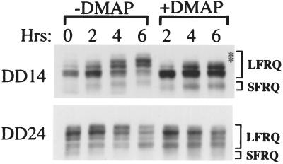
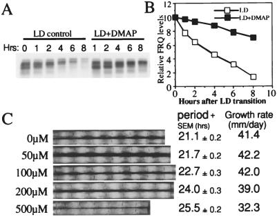
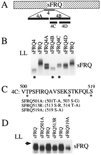
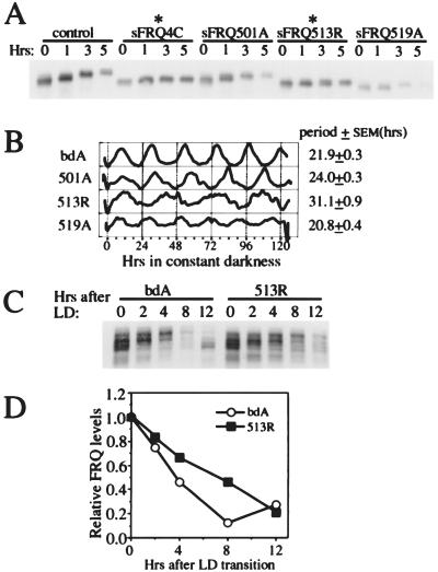
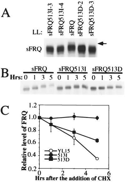
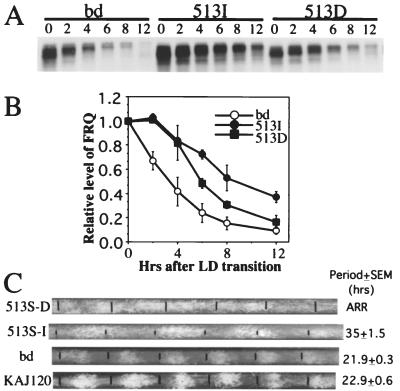
Under free running conditions, FREQUENCY (FRQ) protein, a central component of the Neurospora circadian clock, is progressively phosphorylated, becoming highly phosphorylated before its degradation late in the circadian day. To understand the biological function of FRQ phosphorylation, kinase inhibitors were used to block FRQ phosphorylation in vivo and the effects on FRQ and the clock observed. 6-dimethylaminopurine (a general kinase inhibitor) is able to block FRQ phosphorylation in vivo, reducing the rate of phosphorylation and the degradation of FRQ and lengthening the period of the clock in a dose-dependent manner. To confirm the role of FRQ phosphorylation in this clock effect, phosphorylation sites in FRQ were identified by systematic mutagenesis of the FRQ ORF. The mutation of one phosphorylation site at Ser-513 leads to a dramatic reduction of the rate of FRQ degradation and a very long period (>30 hr) of the clock. Taken together, these data strongly suggest that FRQ phosphorylation triggers its degradation, and the degradation rate of FRQ is a major determining factor for the period length of the Neurospora circadian clock.
Figures
Similar articles
-
Phosphorylation of FREQUENCY protein by casein kinase II is necessary for the function of the Neurospora circadian clock.Mol Cell Biol. 2003 Sep;23(17):6221-8. doi: 10.1128/MCB.23.17.6221-6228.2003. Mol Cell Biol. 2003. PMID: 12917343 Free PMC article.
-
Setting the pace of the Neurospora circadian clock by multiple independent FRQ phosphorylation events.Proc Natl Acad Sci U S A. 2009 Jun 30;106(26):10722-7. doi: 10.1073/pnas.0904898106. Epub 2009 Jun 8. Proc Natl Acad Sci U S A. 2009. PMID: 19506251 Free PMC article.
-
Regulation of the Neurospora circadian clock by casein kinase II.Genes Dev. 2002 Apr 15;16(8):994-1006. doi: 10.1101/gad.965102. Genes Dev. 2002. PMID: 11959847 Free PMC article.
-
Posttranslational regulation of Neurospora circadian clock by CK1a-dependent phosphorylation.Cold Spring Harb Symp Quant Biol. 2007;72:177-83. doi: 10.1101/sqb.2007.72.025. Cold Spring Harb Symp Quant Biol. 2007. PMID: 18419275 Review.
-
The molecular workings of the Neurospora biological clock.Novartis Found Symp. 2003;253:184-98; discussion 102-9, 198-202, 281-4. Novartis Found Symp. 2003. PMID: 14712922 Review.
Cited by
-
Glycogen synthase kinase is a regulator of the circadian clock of Neurospora crassa.J Biol Chem. 2012 Oct 26;287(44):36936-43. doi: 10.1074/jbc.M112.396622. Epub 2012 Sep 6. J Biol Chem. 2012. PMID: 22955278 Free PMC article.
-
Molecular mechanism of suppression of circadian rhythms by a critical stimulus.EMBO J. 2006 Nov 15;25(22):5349-57. doi: 10.1038/sj.emboj.7601397. Epub 2006 Oct 26. EMBO J. 2006. PMID: 17066078 Free PMC article.
-
Phosphorylation of FREQUENCY protein by casein kinase II is necessary for the function of the Neurospora circadian clock.Mol Cell Biol. 2003 Sep;23(17):6221-8. doi: 10.1128/MCB.23.17.6221-6228.2003. Mol Cell Biol. 2003. PMID: 12917343 Free PMC article.
-
A role for protein kinase casein kinase2 α-subunits in the Arabidopsis circadian clock.Plant Physiol. 2011 Nov;157(3):1537-45. doi: 10.1104/pp.111.179846. Epub 2011 Sep 7. Plant Physiol. 2011. PMID: 21900482 Free PMC article.
-
Circadian output, input, and intracellular oscillators: insights into the circadian systems of single cells.Cold Spring Harb Symp Quant Biol. 2007;72:201-14. doi: 10.1101/sqb.2007.72.067. Cold Spring Harb Symp Quant Biol. 2007. PMID: 18419278 Free PMC article. Review.
References
-
- Dunlap J C. Cell. 1999;96:271–290. - PubMed
-
- Young M W. Annu Rev Biochem. 1998;67:135–152. - PubMed
-
- Rosbash M, Allada R, Dembinska M, Guo W Q, Le M, Marrus S, Qian Z, Rutila J, Yaglom J, Zeng H. Cold Spring Harbor Symp Quant Biol. 1996;61:265–278. - PubMed
-
- Hall J C. Trends Neurosci. 1995;18:230–240. - PubMed
-
- Golden S S, Johnson C H, Kondo T. Curr Opin Microbiol. 1998;1:669–673. - PubMed
Publication types
MeSH terms
Substances
Grants and funding
LinkOut - more resources
Full Text Sources
