. 2000 Jan 4;97(1):337-42.
doi: 10.1073/pnas.97.1.337.

Estrogen receptors alpha and beta in the rodent mammary gland

Affiliations

Estrogen receptors alpha and beta in the rodent mammary gland

S Saji et al. Proc Natl Acad Sci U S A. .

Abstract

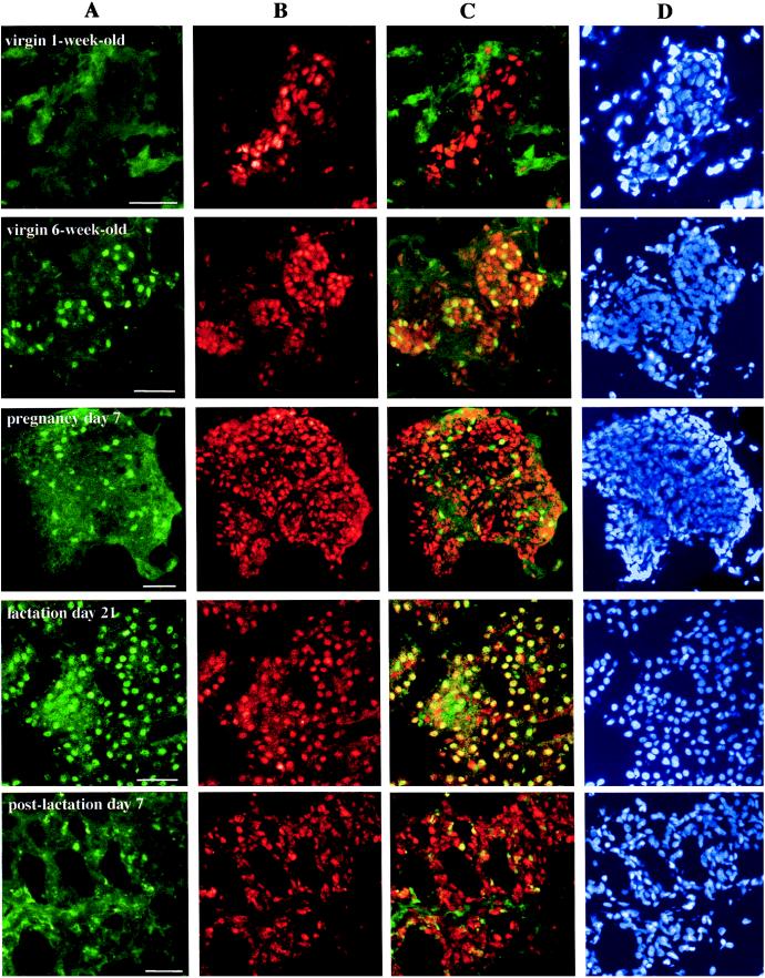
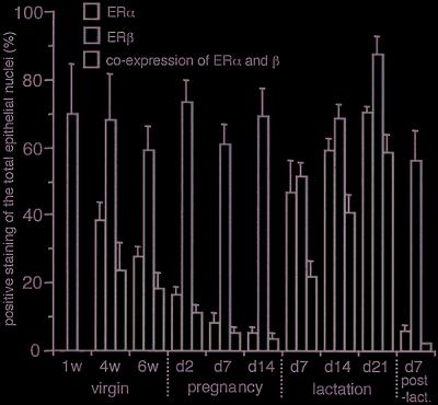
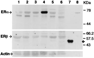
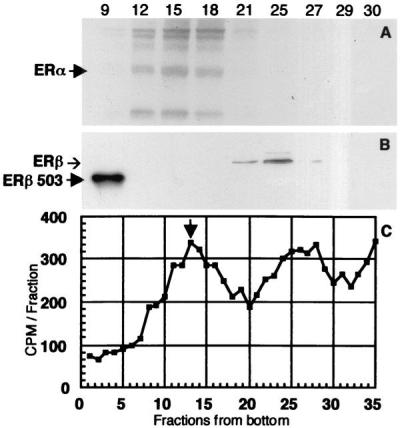
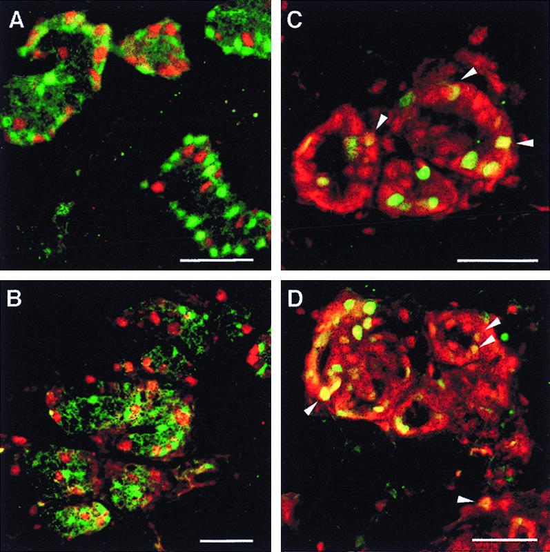
An obligatory role for estrogen in growth, development, and functions of the mammary gland is well established, but the roles of the two estrogen receptors remain unclear. With the use of specific antibodies, it was found that both estrogen receptors, ERalpha and ERbeta, are expressed in the rat mammary gland but the presence and cellular distribution of the two receptors are distinct. In prepubertal rats, ERalpha was detected in 40% of the epithelial cell nuclei. This decreased to 30% at puberty and continued to decrease throughout pregnancy to a low of 5% at day 14. During lactation there was a large induction of ERalpha with up to 70% of the nuclei positive at day 21. Approximately 60-70% of epithelial cells expressed ERbeta at all stages of breast development. Cells coexpressing ERalpha and ERbeta were rare during pregnancy, a proliferative phase, but they represented up to 60% of the epithelial cells during lactation, a postproliferative phase. Western blot analysis and sucrose gradient centrifugation confirmed this pattern of expression. During pregnancy, the proliferating cell nuclear antigen was not expressed in ERalpha-positive cells but was observed in 3-7% of ERbeta-containing cells. Because more than 90% of ERbeta-bearing cells do not proliferate, and 55-70% of the dividing cells have neither ERalpha nor ERbeta, it is clear that the presence of these receptors in epithelial cells is not a prerequisite for estrogen-mediated proliferation.


Figures

Figure 1
Figure 1
Evaluation of the specificity of ERβ 503 antibody. Frozen sections of ventral prostate from 12-week-old male rats were stained by using ERβ 503 antibody (A), ERβ 503 antibody preadsorbed with recombinant ERβ protein (B), and ERβ antibody preadsorbed with BSA (C). (Bars = 50 μm.)
Figure 2
Figure 2
Detection and localization by immunofluorescent labeling of ERα and ERβ proteins in rat mammary glands at different stages of development. Tissue sections from female rats at indicated intervals were stained by sequential exposure of each section to antibodies 6F11, FITC-m, 503, and Cy3-c, followed by DAPI. The green fluorescence spots indicate nuclei containing ERα (A), and the red spots indicate those with ERβ (B). The yellow nuclei observed in overlayered images, especially from lactating glands, indicate that there is coexpression of both receptors in a single epithelial cell (C). Lane D shows the total nuclei in the same section. Pictures are representative of nine slides from three individual rats for each stage. (Bars = 50 μm.)
Figure 3
Figure 3
Detailed analysis of ERα and ERβ expression as indicated by immunofluorescent labeling. Cell nuclei positive for ERα, ERβ, or both were counted and the numbers were expressed as the percentage of total epithelial cell nuclei. Results are mean ± SD of three determinations for each of three individual tissues. Green bars indicate the percentage of cells expressing ERα, red bars indicate those expressing ERβ, and yellow bars indicate those expressing both receptors.
Figure 4
Figure 4
Detection of ERα and ERβ by Western blot analysis. Similar amounts of homogenized tissue lysates were separated by 9% SDS/PAGE and immunoblotted with either ERα antibody 6F11, ERβ antibody LBD, or actin antibody Ab-1. Lanes: 1, virgin 6 week old; 2, pregnancy day 7; 3, pregnancy day 14; 4, lactation day 21; 5, postlactation day 7; 6, ventral prostate; 7, ERβ 503 recombinant protein; 8, ERα recombinant protein. The arrows at the top, middle, and bottom sections indicate ERα, ERβ, and actin proteins, respectively. The bold arrow at the middle section indicates recombinant ERβ 503 protein. The results are representative of triplicate experiments.
Figure 5
Figure 5
(C) Sedimentation analysis of tritiated estradiol binding by receptors in cytosol from lactating rat mammary gland. Arrowhead shows the position of the 8S peak of the receptor present in MCF-7 cell cytosol (ERα). (A and B) Determination of the ERα and ERβ content of representative gradient fractions of both peaks by Western blotting. The arrowhead in A indicates the position of ERα on the blot, and the arrowhead as well as the band in the left lane of B indicate that of ERβ 503.
Figure 6
Figure 6
Colocalization of PCNA with ERα or ERβ. Mammary gland sections from 6-week-old (A and C) and pregnancy day 7 (B and D) female rats were doubly stained with antibodies to PCNA (PC10) and ERα (MC20) (A and B) or to PCNA (PC10) and ERβ (503) (C and D). In A and B, green nuclei indicate cells expressing ERα and red nuclei indicate those containing PCNA. In C and D, green nuclei contain PCNA and red nuclei contain ERβ. The yellow nuclei, some of them marked with arrowheads, indicate coexpression of receptor and PCNA. (Bars = 50 μm.)

References

    1. Russo J, Russo I H. Medicina. 1997;57, Suppl 2:81–91. - PubMed
    1. Prall O W, Rogan E M, Sutherland R L. J Steroid Biochem Mol Biol. 1998;65:169–174. - PubMed
    1. Zeps N, Bentel J M, Papadimitriou J M, D'Antuono M F, Dawkins H J. Differentiation. 1998;62:221–226. - PubMed
    1. Clarke R B, Howell A, Potten C S, Anderson E. Cancer Res. 1997;57:4987–4991. - PubMed
    1. Clarke R B, Howell A, Anderson E. Breast Cancer Res Treat. 1997;45:121–133. - PubMed

Publication types

LinkOut - more resources