

Review
doi: 10.1172/JCI9083.
Lessons learned from the development of an abl tyrosine kinase inhibitor for chronic myelogenous leukemia
Affiliations
- PMID: 10619854
- PMCID: PMC382593
- DOI: 10.1172/JCI9083
Item in Clipboard
Review
Lessons learned from the development of an abl tyrosine kinase inhibitor for chronic myelogenous leukemia
J Clin Invest.
2000 Jan.
No abstract available
Figures
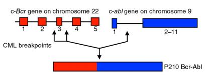
Structure of the Bcr-Abl gene. The Philadelphia chromosome is formed by a reciprocal translocation between chromosomes 9 and 22. Potential breakpoints are indicated by arrows. This resulting translocation replaces the first exon of c-abl with sequences from the Bcr gene.
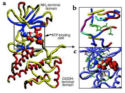
The crystal structure of AMP-PNP bound to Lck was used to made a model of the Abl kinase domain. (a) The general architecture of the Abl kinase domain. The NH2-terminal domain has a 5-strand β-sheet (blue) and 1 α-helix (red). The COOH-terminal domain is made up of several α-helices. ATP is shown in the ATP-binding cleft. (b) Topology of the ATP-binding cleft of the Abl protein kinase. The ATP-binding cleft can be subdivided into 6 regions: adenine-binding region (yellow), hydrophobic region I (blue), hydrophobic region II (aqua), phosphate-binding region (red-orange), the ribose pocket (purple), and the linker region (pink). The ligated ATP is shown in green-blue. (c) Amino acid side chains lining the ATP-binding cleft.
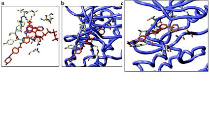
Model of STI 571 bound within the active site of the Abl kinase. (a) Overlay of ATP (red) and STI 571 (orange). Key amino acids that interact with ATP are shown. Binding mode of STI 571 shown from above (b) and from the side (c).
References
-
- Kolibaba KS, Druker BJ. Protein tyrosine kinases and cancer. Biochim Biophys Acta. 1997;1333:F217–F248. - PubMed
-
- Rowley JD. A new consistent abnormality in chronic myelogenous leukaemia identified by quinacrine fluorescence and giemsa staining. Nature. 1973;243:290–293. - PubMed
-
- Heisterkamp N, et al. Localization of the c-abl oncogene adjacent to a translocation break point in chronic myelocytic leukemia. Nature. 1983;306:239–242. - PubMed
-
- Bartram CR, et al. Translocation of c-abl correlates with the presence of a Philadelphia chromosome in chronic myelocytic leukemia. Nature. 1983;306:277–280. - PubMed
-
- Shtivelman E, Lifshitz B, Gale RP, Canaani E. Fused transcript of abl and bcr genes in chronic myelogenous leukaemia. Nature. 1985;315:550–554. - PubMed
Publication types
MeSH terms
Substances
LinkOut - more resources
Full Text Sources
Other Literature Sources
Medical
Miscellaneous
