. 2000 Jan;105(1):79-92.
doi: 10.1172/JCI8066.

Salmonella typhimurium induces epithelial IL-8 expression via Ca(2+)-mediated activation of the NF-kappaB pathway

Affiliations

Salmonella typhimurium induces epithelial IL-8 expression via Ca(2+)-mediated activation of the NF-kappaB pathway

A T Gewirtz et al. J Clin Invest. 2000 Jan.

Abstract

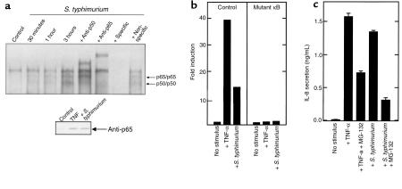
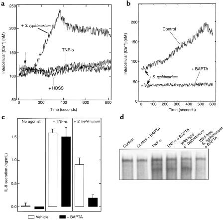
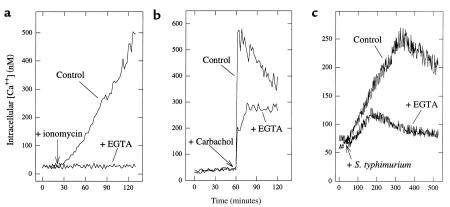
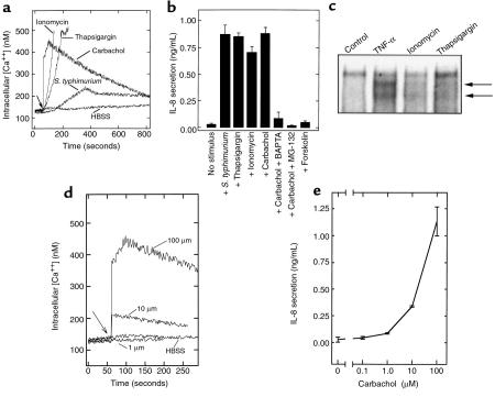
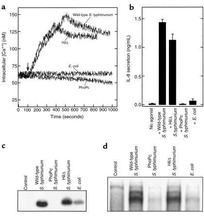
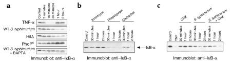
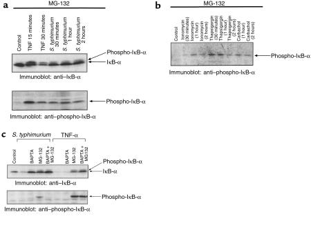
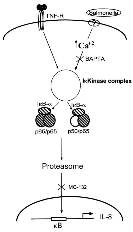
Interactions between the enteric pathogen Salmonella typhimurium and the luminal surface of the intestine provoke an acute inflammatory response, mediated in part by epithelial cell secretion of the chemokine IL-8 and other proinflammatory molecules. This study investigated the mechanism by which this pathogen induces IL-8 secretion in physiologically polarized model intestinal epithelia. IL-8 secretion induced by both the prototypical proinflammatory cytokine TNF-alpha and S. typhimurium was NF-kappaB dependent. However, NF-kappaB activation and IL-8 secretion induced by S. typhimurium, but not by TNF-alpha, was preceded by and required an increase in intracellular [Ca(2+)]. Additionally, agonists that increased intracellular [Ca(2+)] by receptor-dependent (carbachol) or independent (thapsigargin, ionomycin) means also induced IL-8 secretion. Furthermore, the ability of S. typhimurium mutants to induce IkappaB-alpha degradation, NF-kappaB translocation, and IL-8 transcription and secretion correlated precisely with their ability to induce an intracellular [Ca(2+)] increase in model intestinal epithelia, but not with their ability to invade these cells. Finally, S. typhimurium, but not TNF-alpha, induced a Ca(2+)-dependent phosphorylation of IkappaB-alpha. These results indicate that S. typhimurium-induced activation of NF-kappaB-dependent epithelial inflammatory responses proceeds by a Ca(2+)-mediated activation of an IkappaB-alpha kinase. These observations raise the possibility that pharmacologic intervention of the acute inflammatory response can be selectively matched to the specific class of initiating event.


Figures

Figure 1
Figure 1
S. typhimurium and TNF-α induce IL-8 expression by an NF-κB–mediated mechanism. (a) EMSAs with an IL-8 NF-κB motif probe. Nuclear extracts were derived from model intestinal epithelia maintained under control conditions or coincubated with wild-type S. typhimurium as described in Methods for the indicated times. Addition of unlabeled competitor probe or antisera when indicated. Inset: nuclear extracts were Western blotted with anti-p65 as described in Methods. (b) Transient transfection assay with IL-8 reporter constructs. Transfected HT-29 cl19A cells were treated as indicated. Data are reported as fold induction of CAT activity over basal. (c) IL-8 ELISA from model epithelia. Cells were incubated with basolateral TNF-α (10 ng/mL) or apical wild-type S. typhimurium, either with or without 1 hour pretreatment with 50 μM MG-132. Basolateral IL-8 secretion was measured 5 hours later.
Figure 2
Figure 2
S. typhimurium, but not TNF-α, induce Ca2+ mobilization that is required for NF-κB activation and IL-8 secretion. (a, b) Fluorometric assay of intracellular calcium. Model intestinal epithelia were loaded with the Ca2+ indicator Fura-2 and placed in a thermostated spectrofluorometer and intracellular Ca2+ was measured as described in Methods. TNF-α was added basolaterally or S. typhimurium was added apically as indicated. In b, intracellular [Ca2+] was measured in response to S. typhimurium in the absence or presence of intracellular BAPTA, which was simultaneously loaded with Fura-2. (c) IL-8 ELISA. Model intestinal epithelia were stimulated basolaterally with TNF-α or infected apically with S. typhimurium in the presence (filled bars) or absence (open bars) of 15 μM BAPTA. Basolateral IL-8 secretion was measured 5 hours later. (d) EMSA with IL-8 probe. Model intestinal epithelia were treated as in c. After stimulation (30 minutes for TNF-α; 1 hour for S. typhimurium), nuclear extracts were isolated and NF-κB translocation assessed as described in Methods.
Figure 3
Figure 3
Chelation of extracellular Ca2+ indicates S. typhimurium–induced [Ca2+] increase results from both intracellular stores and Ca2+ influx. Fluorometric assays of intracellular calcium. Intracellular [Ca2+] was measured in Fura-2–loaded model intestinal epithelia in response to (a) ionomycin (1 μg/mL), (b) carbachol (100 μM), or (c) S. typhimurium in the presence or absence of EGTA (5 mM), which was added 10 seconds before the addition of an agonist.
Figure 4
Figure 4
Stimuli that induce an increase in intracellular [Ca2+] elicit IL-8 secretion. (a) Fluorometric assays of intracellular [Ca2+]. Intracellular [Ca2+] was measured in Fura-2–loaded model intestinal epithelia in response to S. typhimurium, thapsigargin (10 μM) ionomycin (1 μg/mL), carbachol (100 μM), or forskolin (10 μM). (b) IL-8 ELISA. Five hours after addition of the agonist, basolateral IL-8 secretion was measured by ELISA. BAPTA (added in AM form) and MG-132 were used when indicated as described in Methods. (c) EMSA with IL-8 κB probe. Model epithelia were treated with TNF-α, ionomycin (1 μg/mL) or thapsigargin (10 μM) for 1 hour before extract preparation. NF-κB complexes are indicated with arrows. (d) Intracellular [Ca2+] and (e) IL-8 secretion, in response to the indicated dose of carbachol.
Figure 5
Figure 5
S. typhimurium ability to induce an increase in [Ca2+] correlates with its ability to elicit activation of NF-κB and subsequent IL-8 secretion. Model intestinal epithelia were treated with the indicated strain of S. typhimurium or a nonpathogenic strain of gut E. coli. Bacteria were applied apically. (a) Fluorometric assay of intracellular [Ca2+]. (b) IL-8 secretion measured by ELISA. IL-8 was assayed from the basolateral media after 5 hours. (c) Northern blot probed with IL-8. T84 cell mRNA prepared after infection for 5 hours with the indicated organisms. (d) EMSA with IL-8 probe. Nuclear extracts prepared 3 hours after infection.
Figure 6
Figure 6
Proinflammatory S. typhimurium induce Ca2+-dependent degradation of IκB-α. Immunoblots with anti–IκB-α antibodies. (a) Whole cell extracts were prepared at the indicated times from model intestinal epithelia treated with basolateral TNF-α, apical wild-type S. typhimurium (WT), HilΔ and PhoPc mutants, and wild type with BAPTA-pretreated epithelia. (b) Monolayers were treated with basolateral ionomycin (1 μg/mL), thapsigargin (10 μM), or carbachol (100 μM) for the times indicated. (c) Model epithelia were treated with 20 μg/mL cycloheximide, S. typhimurium, or CHX 1 hour before S. typhimurium infection.
Figure 7
Figure 7
Proinflammatory S. typhimurium induces Ca2+-dependent phosphorylation of IκB-α. Immunoblots with anti–IκB-α or anti–phospho-IκB-α antibodies. (a) All model epithelia were pretreated with 50 μM MG-132 for 30 minutes. Whole cell extracts were prepared at the indicated times from cells treated with basolateral TNF-α or apical wild-type S. typhimurium (WT). Extracts were electrophoresed and immunoblotted with anti–IκB-α and anti–phospho-IκB-α antibodies. (b) Cells were pretreated with 50 μM MG-132 for 1 hour before addition of agonists as described in Figure 6b before immunoblot with antiphospho-IκB-α antisera. (c) Epithelia were treated with MG-132 and/or BAPTA as described in Methods, before activation with TNF-α (30 minutes) or Salmonella (1 hour). Extracts were immunoblotted with anti–IκB-α and anti–phospho-IκB-α antibodies as indicated.
Figure 8
Figure 8
Model of S. typhimurium–induced NF-κB activation. Both TNF-α and S. typhimurium induce NF-κB nuclear translocation via activation of an IκB kinase and subsequent proteasomal degradation of IκB-α. However, S. typhimurium–mediated, but not TNF-α–mediated, activation of the kinase requires an increase in cytosolic [Ca2+].

Comment in

References

    1. Madara JL. Pathobiology of the intestinal epithelial barrier. Am J Pathol. 1990;137:1273–1281. - PMC - PubMed
    1. Yamada, T., Alpers, D.H., Owyang, C., Powell, D.W., and Silverstein, F.E. 1995. Textbook of gastroenterology. J.B. Lippincott. Philadelphia, PA. 1606–1629.
    1. Takeuchi A. Electron microscope studies of experimental Salmonella infection. Am J Pathol. 1967;50:109–119. - PMC - PubMed
    1. McCormick B, Gewirtz A, Madara JL. Epithelial crosstalk with bacteria and immune cells. Curr Opin Gastro. 1998;14:492–497.
    1. McCormick BA, Miller SI, Madara JL. New insights on molecular pathways utilized by salmonella species in cell binding. Front Biosci. 1996;1:d131–d145. - PubMed

Publication types

MeSH terms