. 2000 Jan;105(1):101-11.
doi: 10.1172/JCI7954.

Chemotaxis of primitive hematopoietic cells in response to stromal cell-derived factor-1

Affiliations

Chemotaxis of primitive hematopoietic cells in response to stromal cell-derived factor-1

D Y Jo et al. J Clin Invest. 2000 Jan.

Abstract

Stromal cell-derived factor-1 (SDF-1) provides a potent chemotactic stimulus for CD34(+) hematopoietic cells. We cultured mobilized peripheral blood (PB) and umbilical cord blood (CB) for up to 5 weeks and examined the migratory activity of cobblestone area-forming cells (CAFCs) and long-term culture-initiating cells (LTC-ICs) in a transwell assay. In this system, SDF-1 or MS-5 marrow stromal cells placed in the lower chamber induced transmembrane and transendothelial migration by 2- and 5-week-old CAFCs and LTC-ICs in 3 hours. Transmigration was blocked by preincubation of input CD34(+) cells with antibody to CXCR4. Transendothelial migration of CB CAFCs and LTC-ICs was higher than that of PB. We expanded CD34(+) cells from CB in serum-free medium with thrombopoietin, flk-2 ligand, and c-kit ligand, with or without IL-3 and found that CAFCs cultured in the absence of IL-3 had a chemotactic response equivalent to noncultured cells, even after 5 weeks. However, addition of IL-3 to the culture reduced this response by 20-50%. These data indicate that SDF-1 induces chemotaxis of primitive hematopoietic cells signaling through CXCR4 and that the chemoattraction could be downmodulated by culture ex vivo.


Figures
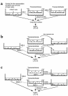
Figure 1
Figure 1
(a) Experimental design for “closed system” studies of transmembrane (middle) and transendothelial (right) chemotaxis of CAFCs and LTC-ICs in CD34+ populations placed in the upper chamber of a transwell system, with or without a monolayer of endothelial cells (BMEC-1), with MS-5 cells in the lower chamber as a source of SDF-1 and as a support for CAFCs and LTC-ICs in long-term (5-week) cultures. (b) Experimental design for “open system” studies transmembrane and transendothelial chemotaxis of CAFCs and LTC-ICs in CD34+ populations placed in the upper chamber of a transwell system, with or without a monolayer of endothelial cells (BMEC-1), and with recombinant SDF-1 in the lower chamber (middle). CAFCs and LTC-ICs are assayed by recovery of cells from the upper and lower chambers and secondary transfer to MS-5 monolayers for long-term (5-week) cultures (right). (c) Design as in b but substituting an established monolayer of MS-5 in the lower chamber as a source of SDF-1.
Figure 2
Figure 2
Transmembrane migration of LTC-ICs to an SDF-1 gradient. SDF-1 (100 ng/mL) was added in the lower chambers of 24-well transwell plates (Figure 1b), with 1 × 10⁴ CB CD34+ cells in a upper chamber. At indicated time points, the upper chambers were removed and the migrated cells and nonmigrated cells were transferred onto separate MS-5 feeder layers in 6-well plates for LTC.
Figure 3
Figure 3
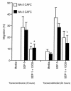
Transmigration of week-2 and week-5 CAFC induced by SDF-1 and its blocking by mAb to CXCR4 (12G5). SDF-1 (100 ng/mL) was added in the lower chambers of 6-well plates, and CB CD34+ cells were added to the upper chambers (3 μm) with or without a BMEC-1 monolayer. For blocking the migration, CD34+ cells were preincubated with 12G5 for 30 minutes. After 3 hours (transmembrane migration) and 24 hours (transendothelial migration), the migrated cells were transferred onto MS-5 feeder layers in 6-well plates and LTC was performed. The percent of migration of CAFCs was calculated based on the numbers in control populations, in which CD34+ cells were put directly onto MS-5 feeder layers. Data are mean ± SD of triplicate experiment. *P < 0.05 compared with matched, 12G5 no-treated transmigration to an SDF-1 gradient (i.e., SDF-1 versus SDF-1 + 12G5).
Figure 4
Figure 4
Transmembrane migration of week-2 CAFCs (a), week-5 CAFCs (b), and LTC-ICs (c) from mobilized PB and CB. CD34+ cells (5 × 10³ cells) were added to the upper chambers of 6-well transwell plates, and the upper chambers were placed in 6-well plates containing an MS-5 feeder layer that had been incubated for 3 days. After incubation of the plates at 37°C during indicated periods, the upper chambers were removed. After 2 weeks and 5 weeks of culture of the lower chambers containing migrated cells, CAFCs were scored and LTC-ICs were enumerated. The numbers of week-2 CAFCs from 5,000 CD34+ cells were 45.3 ± 23.2 and 96.3 ± 108.5 for PB and CB, respectively. The numbers of week-5 CAFC from 5,000 CD34+ cells were 135.5 ± 42.4 and 162.5 ± 88.3 for PB and CB, respectively. The numbers of LTC-ICs from 5,000 CD34+ cells were 58.4 ± 38.3 and 71.2 ± 60.5 for PB and CB, respectively. Data are mean ± SD of 3 or more independent experiments (filled triangle, PB with MS-5 in the lower chamber; filled circle, CB with MS-5 in the lower chamber; open triangle, PB with medium in the lower chamber; open circle, CB with medium in the lower chamber).
Figure 5
Figure 5
Transendothelial migration of week-2 CAFCs (a), week-5 CAFCs (b), and LTC-ICs (c) from mobilized PB and CB. CD34+ cells (5 × 10³ cells) were added to the upper chambers of 6-well transwell plates coated with confluent BMEC-1 monolayer, and the upper chambers were placed in 6-well plates containing an MS-5 feeder layer that had been incubated for 3 days. After incubation of the plates at 37°C for indicated periods, the upper chambers were removed and LTC was performed. The numbers of week-2 CAFCs from 5,000 CD34+ cells were 45.3 ± 23.2 and 96.3 ± 108.5 for PB and CB, respectively. The number of week-5 CAFCs from 5,000 CD34+ cells were 135.5 ± 42.4 and 162.5 ± 88.3 for PB and CB, respectively. The numbers of LTC-ICs from 5,000 CD34+ cells were 58.4 ± 38.3 and 71.2 ± 60.5 for PB and CB, respectively. Data are mean ± SD of 3 or more independent experiments (filled triangle, PB with MS-5 in the lower chamber; filled circle, CB with MS-5 in the lower chamber; open triangle, PB with medium in the lower chamber; open circle, CB with medium in the lower chamber). *P < 0.05 compared with PB at each time point.
Figure 6
Figure 6
The correlation between the percent of migration of week-5 CAFCs and percent of migration of LTC-ICs in transmembrane and transendothelial migration experiments.
Figure 7
Figure 7
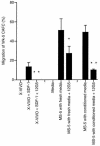
Blocking of the transmembrane migration of CAFCs and LTC-ICs toward MS-5 by neutralizing mAb to CXCR4 (12G5). CB CD34+ cells, which were preincubated with 12G5 for 30 minutes, were allowed to migrate across the 3-μm microporous membrane (6-well transwell plates) toward MS-5 for 3 hours. After removal of the upper chambers, LTC was performed. CAFC was scored at week 2 and week 5, and LTC-ICs were enumerated. The percent of migration of CAFCs and LTC-ICs was calculated from the numbers in input controls, in which CD34+ cells were put directly onto MS-5 feeder layers. Data are mean ± SD of quadruplicate experiments. *P < 0.01; **P < 0.001 compared with matched, 12G5 nontreated transmigration to a SDF-1 or conditioned media gradient.
Figure 8
Figure 8
Blocking of the transendothelial migration of week-5 CAFCs toward MS-5 or an SDF-1 gradient by mAb to CXCR4 (12G5). PB CD34+ cells, which were preincubated with 12G5 for 30 minutes, were allowed to migrate across 3-μm membrane (24-well transwell) coated with confluent BMEC-1 monolayers toward MS-5 with conditioned media or fresh media replaced just before the experiment, or SDF-1 (100 ng/mL in lower chamber containing X-VIVO) for 24 hours. Cells that migrated into the lower chamber were transferred onto MS-5 feeder layers, and LTC was performed. Data are mean ± SD of triplicate experiments. *P < 0.05; **P < 0.01 compared with matched, 12G5 nontreated transmigration to a SDF-1 or conditioned media gradient.
Figure 9
Figure 9
Ex vivo expansion of CB CD34+ cells and transmigration of the expanded cells. CB CD34+ cells (1 × 10⁴ cells) were cultured with various cytokine combinations of TPO, KL, FL, and IL-3 in serum-free medium QBSF-60 (Quality Biologicals, Gaithersburg, Maryland, USA) for 7 days and 14 days. The expanded cells from 0.5 × 10³ to 1 × 10³ CD34+ cells were added to the upper chambers of 6-well plates and allowed to migrate across the 3-μm membrane toward MS-5. (a) Expansion of total colony-forming cells (CFCs). The number of colonies per 1,000 CD34+ cells was 168.6 ± 32.1. (b) Expansion of week-5 CAFCs. The number of week-5 CAFC from 5,000 CD34+ cells was 171.6 ± 94.0. (c) Three-hour migration of 7- and 14-day expanded week-5 CAFCs. (d) Twenty-four-hour migration of 7- and 14-day expanded week-5 CAFCs. *P < 0.05 compared with the matched, IL-3 noncontaining cytokine combinations (i.e., TPO + FL versus TPO + FL + KL; TPO + FL + KL versus TPO + FL + KL + IL-3; t test for paired samples). Data are mean ± SD of 4 or more experiments.
Figure 10
Figure 10
Flow cytometry of 7-day expanded cells before expansion (a); expanded cells with TPO + FL (b); expanded cells with TPO + FL + KL (c); expanded cells with TPO + FL + KL + IL-3 (d).
Figure 11
Figure 11
Transendothelial migration of ex vivo expanded CB CD34+ cells for 4 weeks. Cultured cells were examined for 24-hour transendothelial migration of week-2 CAFCs and week-5 CAFCs.

References

    1. Hendrikx PJ, Martens ACM, Hagenbeek A, Keij JF, Visser JWM. Homing of fluorescently labeled murine hematopoietic stem cells. Exp Hematol. 1996;24:129–140. - PubMed
    1. van der Loo JCM, Ploemacher RE. Marrow- and spleen-seeding efficiencies of all murine hematopoietic stem cell subsets are decreased by preincubation with hematopoietic growth factors. Blood. 1995;85:2598–2606. - PubMed
    1. Papayannopoulou T, Craddock C. Homing and trafficking of hemopoietic progenitor cells. Acta Haematol. 1997;97:97–104. - PubMed
    1. Papayannopoulou T, Nakamoto B. Peripheralization of hematopoietic progenitors in primates treated with anti-VLA4 integrin. Proc Natl Acad Sci USA. 1993;90:9374–9378. - PMC - PubMed
    1. Vermeulen M, et al. Role of adhesion molecules in the homing and mobilization of murine hematopoietic stem and progenitor cells. Blood. 1998;92:894–900. - PubMed

Publication types