. 1999 Dec;14(12):2015-26.
doi: 10.1359/jbmr.1999.14.12.2015.

Alkaline phosphatase knock-out mice recapitulate the metabolic and skeletal defects of infantile hypophosphatasia

Affiliations

Alkaline phosphatase knock-out mice recapitulate the metabolic and skeletal defects of infantile hypophosphatasia

K N Fedde et al. J Bone Miner Res. 1999 Dec.

Abstract

Hypophosphatasia is an inborn error of metabolism characterized by deficient activity of the tissue-nonspecific isoenzyme of alkaline phosphatase (TNSALP) and skeletal disease due to impaired mineralization of cartilage and bone matrix. We investigated two independently generated TNSALP gene knock-out mouse strains as potential models for hypophosphatasia. Homozygous mice (-/-) had < 1% of wild-type plasma TNSALP activity; heterozygotes had the predicted mean of approximately 50%. Phosphoethanolamine, inorganic pyrophosphate, and pyridoxal 5'-phosphate are putative natural substrates for TNSALP and all were increased endogenously in the knock-out mice. Skeletal disease first appeared radiographically at approximately 10 days of age and featured worsening rachitic changes, osteopenia, and fracture. Histologic studies revealed developmental arrest of chondrocyte differentiation in epiphyses and in growth plates with diminished or absent hypertrophic zones. Progressive osteoidosis from defective skeletal matrix mineralization was noted but not associated with features of secondary hyperparathyroidism. Plasma and urine calcium and phosphate levels were unremarkable. Our findings demonstrate that TNSALP knock-out mice are a good model for the infantile form of hypophosphatasia and provide compelling evidence for an important role for TNSALP in postnatal development and mineralization of the murine skeleton.


Figures

FIG. 1
FIG. 1
Plasma ALP activity determined < 24 h after birth. PCR analyses previously demonstrated(–10) that genotype is the basis for the three clusters of ALP activity reflecting knock-out (−/−), heterozygote (+/−), and control (+/+) mice. Individual values, means (circle in a square, circle in a circle), and SEM are depicted.
FIG. 2
FIG. 2
Body weight versus age in EM versus LJ mice. Knock-out mice have lighter body weights despite rescue with soft diets and PL treatment (see Materials and Methods). The weight deficiency is greater for the EM knockouts.
FIG. 3
FIG. 3
Substrate accumulation. Marked elevations of the TNSALP substrates found in hypophosphatasia patients occur in the EM and LJ knock-out mice (note log scale). EM mice were examined at 25 days of age for levels of urinary PPi (+/+, n = 6; −/−, n = 5), and PEA (+/+, n = 5; −/−, n = 7). Plasma PLP and PL data are shown for 10-to 14-day-old mice (+/+, n = 13; −/−, n = 9) as previously reported.(8) LJ mice were examined at 6 days of age for levels of urinary PPi (+/+, n = 14; −/−, n = 8), urinary PEA (+/+, n = 6; −/−, n = 7), and plasma PLP and PL (+/+, n = 6; −/−, n = 4). Urinary data were normalized to grams of creatinine (Crt).
FIG. 4
FIG. 4
Radiographs of the lower quadrants or of the knees. Findings are shown at the indicated ages. (A, B, E, and F) EM mice. (C and D) LJ mice. Although knock-out mice appear normal at 6 days of age, delayed appearance of epiphyses or secondary ossification centers (white arrows), fractures (open arrow), osteopenia, and bone deformities are noted with aging.
FIG. 5
FIG. 5
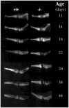
Detailed radiographs of the hind limbs of LJ mice. There is delayed appearance of secondary ossification centers and frayed metaphyses in the knock-out mice at the ages indicated.
FIG. 6
FIG. 6
Histology of proximal tibias of 1-day-old EM mice. (A) Toluidine staining. Epiphyses (E) containing undifferentiated chondrocytes, growth plates (GP), and bone (B). Development is similar in control and knock-out mice. (B) ALP histochemistry (purple) and mineral staining (alizarin red). Normal mineralization at this early age is seen in the knock-out mouse, despite the complete absence of ALP staining. (C) ALP histochemistry and mineral staining at higher magnification. The +/+ control shows characteristic ALP staining which is nearly absent in the resting zone (RZ), but gradually increases in intensity in the proliferative zone (PZ) and the hypertrophic zone (HZ). Strong staining of osteoblasts and periosteal cells is also noted. No ALP staining is observed in the knock-out. In both mice, red-staining mineral is observed between the columns of hypertrophic cells (mineralization front) and was observed in the cortical bone.
FIG. 7
FIG. 7
Histology of older mice. (A) At 18 days of age, toluidine-stained proximal tibial growth plate of LJ mice. In the −/− knock-out mouse, a blunted lower hypertrophic zone with widened vascular invasion channels is seen; the lower hypertrophic zone chondrocytes are nested. (B) At 25 days-of-age, modified Masson trichrome-stained proximal tibial growth plate of EM mice. In the knock-out, there is an enlarged upper hypertrophic zone (UHZ); the lower hypertrophic zone (LHZ) is blunted, and blue-staining mineral aberrantly extends between the columns of chondrocytes. (C) At 25 days of age, modified Masson trichrome-stained diaphyseal shaft of EM mice. The diaphysis of the knock-out mouse shows a widened osteoid seam (pink-staining: O) that extends into the blue-staining mineralized bone. (D) At 101 days of age, modified Masson trichrome stain of trabecular bone tissue of EM mice. Gross osteoidosis (pink-staining material: O) is seen in the knock-out mouse.

Similar articles

Cited by

References

    1. Whyte MP. Hypophosphatasia. In: Scriver CR, Beaudet AL, Sly WS, Valle D, editors. The Metabolic and Molecular Bases of Inherited Disease. 8. McGraw-Hill; New York, NY, U.S.A: in press.
    1. Whyte MP. Hypophosphatasia and the role of alkaline phosphatase in skeletal mineralization. Endocr Rev. 1994;15:439–461. - PubMed
    1. Zurutuza L, Muller F, Gibrat JF, Traillandier A, Simon-Bouy B, Serre JL, Mornet E. Correlations of genotype and phenotype in hypophosphatasia. Hum Mol Genet. 1999;8:1039–1046. - PubMed
    1. Whyte MP. Hypophosphatasia. In: Econs MC, editor. The Genetics of Osteoporosis and Metabolic Bone Disease. Humana Press, Inc; Totowa, NJ, U.S.A: in press.
    1. Fraser D. Hypophosphatasia. Am J Med. 1957;22:730–746. - PubMed

Publication types

MeSH terms