. 2000 Jan;74(2):834-45.
doi: 10.1128/jvi.74.2.834-845.2000.

Role of pseudorabies virus Us9, a type II membrane protein, in infection of tissue culture cells and the rat nervous system

Affiliations

Role of pseudorabies virus Us9, a type II membrane protein, in infection of tissue culture cells and the rat nervous system

A D Brideau et al. J Virol. 2000 Jan.

Abstract

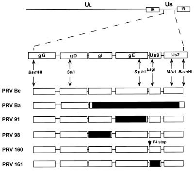
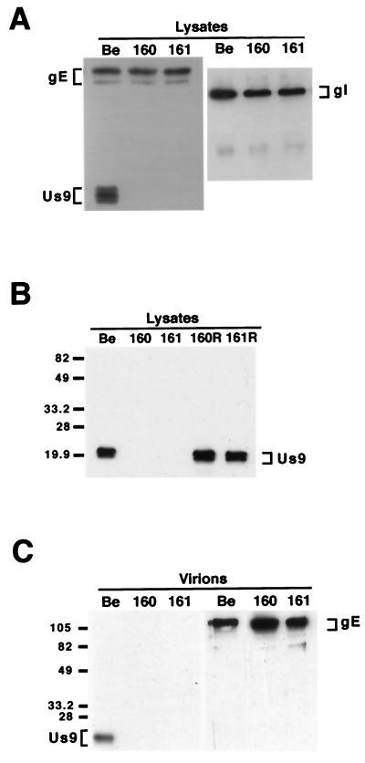
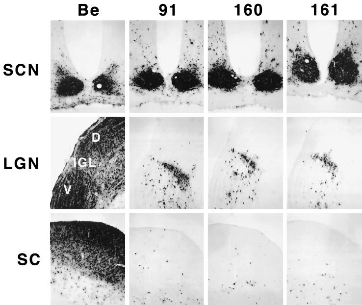
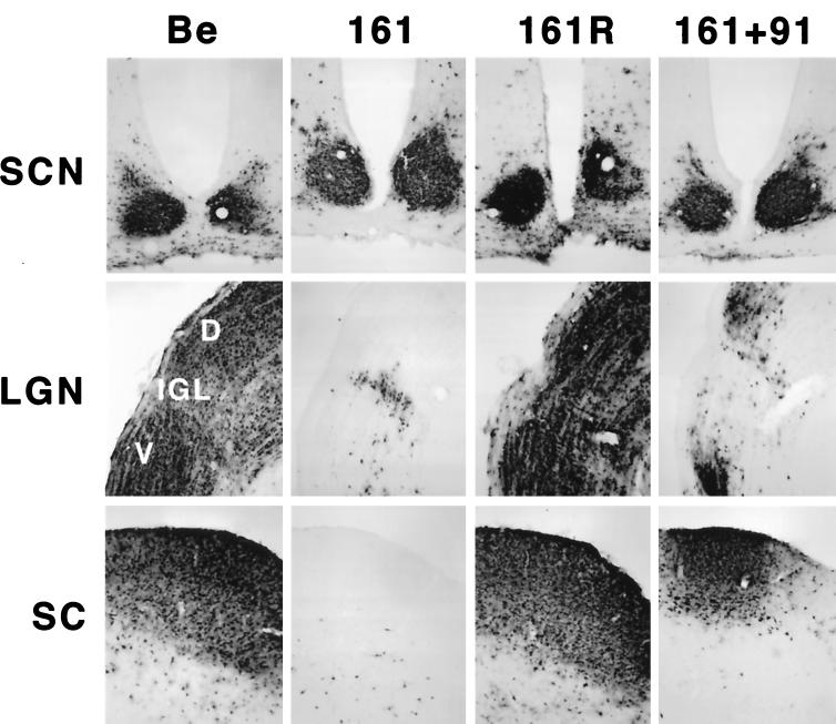
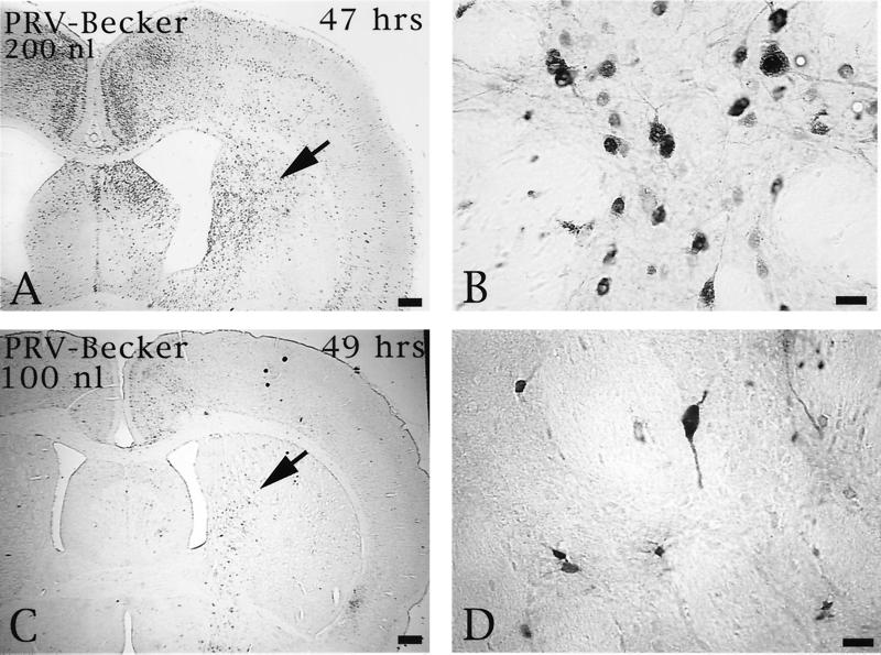
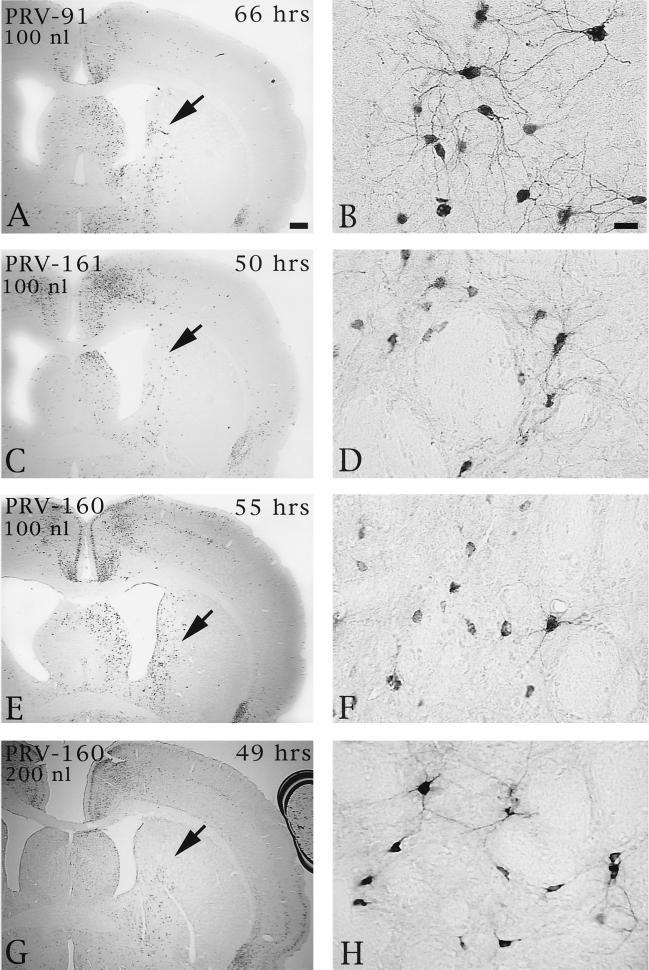
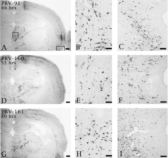
The protein product of the pseudorabies virus (PRV) Us9 gene is a phosphorylated, type II membrane protein that is inserted into virion envelopes and accumulates in the trans-Golgi network. It is among a linked group of three envelope protein genes in the unique short region of the PRV genome which are absent from the attenuated Bartha strain. We found that two different Us9 null mutants exhibited no obvious phenotype after infection of PK15 cells in culture. Unlike those of gE and gI null mutants, the plaque size of Us9 null mutants on Madin-Darby bovine kidney cells was indistinguishable from that of wild-type virus. However, both of the Us9 null mutants exhibited a defect in anterograde spread in the visual and cortical circuitry of the rat. The visual system defect was characterized by restricted infection of a functionally distinct subset of visual projections involved in the temporal organization of behavior, whereas decreased anterograde spread of virus to the cortical projection targets was characteristic of animals receiving direct injections of virus into the cortex. Spread of virus through retrograde pathways in the brain was not compromised by a Us9 deletion. The virulence of the Us9 null mutants, as measured by time to death and appearance of symptoms of infection, also was reduced after their injection into the eye, but not after cortical injection. Through sequence analysis, construction of revertants, measurement of gE and gI protein synthesis in the Us9 null mutants, and mixed-infection studies of rats, we conclude that the restricted-spread phenotype after infection of the rat nervous system reflects the loss of Us9 and is not an indirect effect of the Us9 mutations on expression of glycoproteins gE and gI. Therefore, at least three viral envelope proteins, Us9, gE, and gI, function together to promote efficient anterograde transneuronal infection by PRV in the rat central nervous system.


Figures

FIG. 1
FIG. 1
PRV genome and map of virus strains used in this study. The BamHI 7 fragment of the unique short (Us) region is expanded to show the genes contained in this fragment. The Us deletion of PRV Bartha (Ba) is indicated by a black box. PRV 91 contains a deletion removing the gE coding sequences. PRV 98 is a gI null virus. PRV 160 contains a nonsense stop mutation (stop) at position 4 in place of a phenylalanine residue (F) in the Us9 open reading frame. PRV 161 contains a 258-bp deletion in the Us9 open reading frame. Relevant restriction enzyme sites used in construction of the various recombinant viruses are indicated. Ul, unique long region; PRV Be, PRV Becker; IR, internal repeat.
FIG. 2
FIG. 2
Western blot analysis of Us9 null viruses and revertants. Monolayers of PK15 cells were infected with PRV Becker, PRV 160, and PRV 161 (A and B) and the revertant viruses PRV 160R and PRV 161R (B) at an MOI of 10. Cellular extracts were prepared after 15 h of infection, and approximately 10 μg of total cell lysate was fractionated on an SDS–12.5% polyacrylamide gel, transferred to nitrocellulose, and analyzed by Western blotting with gE, gI, or Us9 antiserum. (C) Viral particles were isolated from the medium of PK15 cells infected for 15 h with either PRV Becker, PRV 160, or PRV 161 (MOI = 10) by centrifugation through sucrose. Virions were analyzed by Western blotting with either gE or Us9 antiserum. The migration of molecular mass markers is indicated on the left in kilodaltons.
FIG. 3
FIG. 3
Anterograde transport of Us9 null viruses in the rodent visual system. Approximately 2.5 × 10⁵ to 2 × 10⁶ PFU of PRV Becker (Be), PRV 91, PRV 160, or PRV 161 was injected into the vitreous humor of Sprague-Dawley male rats. At the time of imminent death, the animals were sacrificed, and the brains were fixed and sliced into 35-μm-thick coronal sections with a freezing microtome. Viral antigen was detected with rabbit polyvalent antiserum Rb133. Representative sections are shown for each virus. LGN, lateral geniculate nuclei; D, dorsal; V, ventral.
FIG. 4
FIG. 4
Reversion and complementation analysis of the Us9 null spread defect. The spread patterns of virus following intraocular injection of animals with PRV Becker (Be), PRV 161, PRV 161R, and a 1:1 (vol/vol) mixed population of PRV 161 and PRV 91 are shown. The infected tissue was processed and analyzed as described in the legend for Fig. 3. Abbreviations are as used for Fig. 3.
FIG. 5
FIG. 5
The extent of anterograde transneuronal infection of the striatum approximately 48 h after injection of 100 (C and D) or 200 nl (A and B) of PRV Becker into the PFC. The areas of the striatum indicated by the arrows in panels A and C are shown at higher magnification in panels B and D, respectively. Note that the extent of anterograde transneuronal infection of the striatum was dramatically increased by injection of the higher concentration of virus. Bars: A and C, 550 μm; B and D, 20 μm.
FIG. 6
FIG. 6
The extent of anterograde transneuronal infection of striatal neurons following injection of PRV 91 (A and B), PRV 161 (C and D), or PRV 160 (E and F) into the medial PFC. The areas of the striatum indicated by the arrows in panels A, C, E, and G are shown at higher magnification in panels B, D, F, and H, respectively. Note that the relative magnitudes of striatal infection produced by injection of 100 nl of PRV 160 or PRV 161 into PFC are similar at 50 to 55 h following injection (compare panels C and D with E and F). In contrast, 66 h was required to achieve a similar magnitude of anterograde infection after injection of PRV 91. It is also important to note that injection of 100 (E and F) or 200 nl (G and H) of PRV 160 into the PFC produced similar magnitudes of anterograde transneuronal infection in the striatum. This contrasted with the concentration-dependent differences in the extent of infection produced by PRV Becker (Fig. 5). Bar in A, 500 μm (same scale for panels C, E, and G); bar in B, 20 μm (same scale for panels D, F, and H).
FIG. 7
FIG. 7
Extent of retrograde infection of thalamus and perirhinal cortex following injection of PRV 91 (A to C), PRV 160 (D to F), or PRV 161 (G to I) into PFC. The boxed areas shown in panel A designate the regions of the thalamus and perirhinal cortex shown at higher magnification in the photomicrographs to the right in each row (thalamus in panels B, E, and H; perirhinal cortex in panels C, F, and I). Note that the levels of retrograde infection produced by all three viruses are similar even though the animal infected with PRV 91 survived 66 h and those injected with PRV 160 and PRV 161 survived 55 and 50 h, respectively. Bars for A, D, and G, 500 μm; bars for B, E, and H, 50 μm; bars for C, F, and I, 100 μm.

References

    1. Babic N, Klupp B, Brack A, Mettenleiter T C, Ugolini G, Flamand A. Deletion of glycoprotein gE reduces the propagation of pseudorabies virus in the nervous system of mice after intranasal inoculation. Virology. 1996;219:279–284. - PubMed
    1. Balan P, Davis-Poynter N, Bell S, Atkinson H, Browne H, Minson T. An analysis of the in vitro and in vivo phenotypes of mutants of herpes simplex virus type 1 lacking glycoproteins gG, gE, gI or the putative gJ. J Gen Virol. 1994;75:1245–1258. - PubMed
    1. Banfield B W, Yap G S, Knapp A C, Enquist L W. A chicken embryo eye model for the analysis of alphaherpesvirus neuronal spread and virulence. J Virol. 1998;72:4580–4588. - PMC - PubMed
    1. Ben-Porat T, Kaplan A S. Molecular biology of pseudorabies virus. In: Roizman B, editor. The herpesviruses. New York, N.Y: Plenum Publishing Corp.; 1985. pp. 105–173.
    1. Brideau A D, Banfield B W, Enquist L W. The Us9 gene product of pseudorabies virus, an alphaherpesvirus, is a phosphorylated, tail-anchored type II membrane protein. J Virol. 1998;72:4560–4570. - PMC - PubMed

Publication types

MeSH terms

Substances

LinkOut - more resources