

Increased serum concentrations of soluble HLA-class I antigens in hepatitis C virus related mixed cryoglobulinaemia
- PMID: 10627422
- PMCID: PMC1752989
- DOI: 10.1136/ard.59.1.20
Increased serum concentrations of soluble HLA-class I antigens in hepatitis C virus related mixed cryoglobulinaemia
Abstract
Objective: To investigate whether quantitative alterations of both beta(2)microglobulin (beta(2)micro) associated HLA class I heavy chains (sHLA-I) and beta(2) micro free class I heavy chains (sHLA-FHC) in sera of patients with hepatitis C virus (HCV) infection occur and whether they distinguish patients with mixed cryoglobulinaemia (MC).
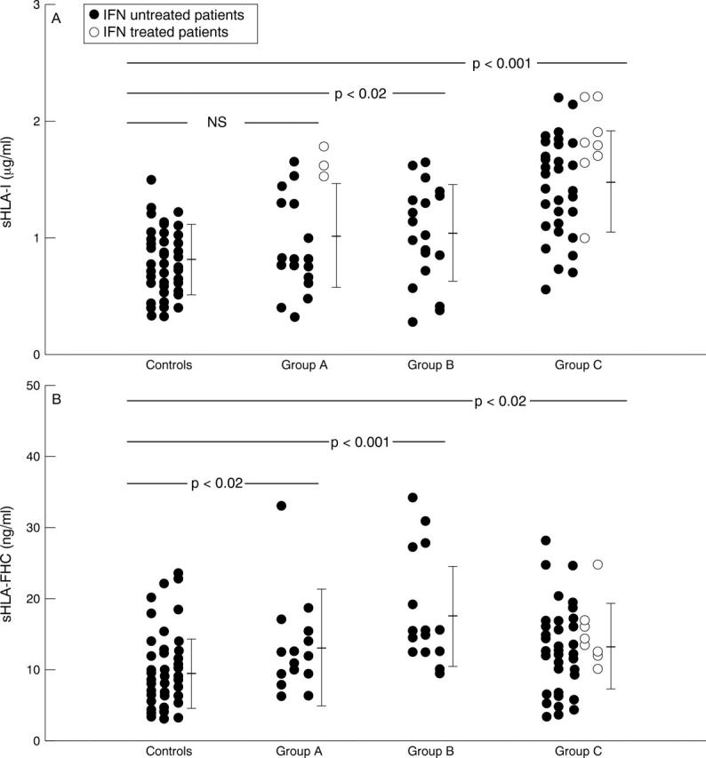
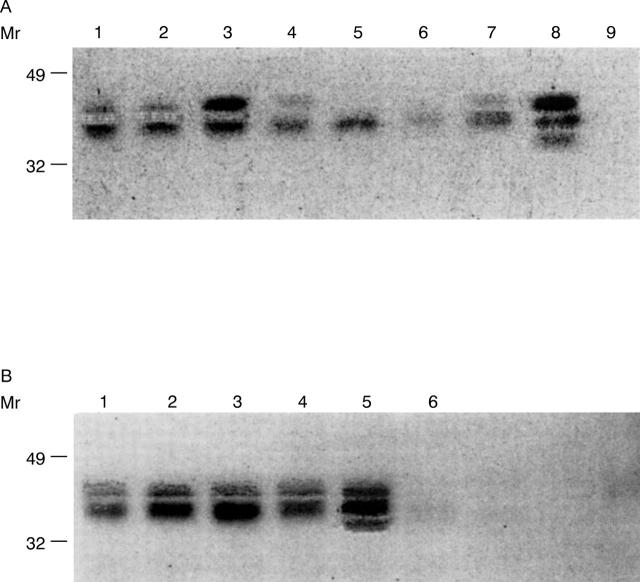
Methods: 83 HCV infected patients were studied and divided into three groups: (A) without cryoglobulinaemia (n=21), (B) with polyclonal MC (n=20), (C) with monoclonal MC (n=42). Serum sHLA-I and sHLA-FHC were measured by double determinant radioimmunoassay using monoclonal antibodies: TP25.99 as catching antibody, and NAMB-1 and HC-10 as revealing antibodies. Western blot identified HLA-I isoforms.
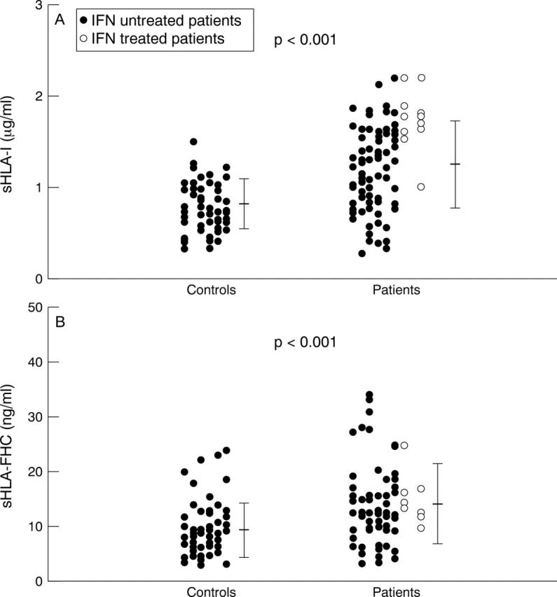
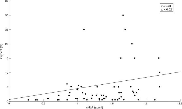
Results: The serum concentrations of sHLA-I and of sHLA-FHC in HCV infected patients versus controls were respectively 1.3(0.5) microg/ml (mean (SD)) versus 0.8 (0.3) (p<0. 001) and 13.9 (7.1) ng/ml versus 9.2 (5) (p<0.001). sHLA-I were 1.01 (0.4) microg/ml in group A, 1.04 (0.4) microg/ml in group B, and 1. 47 (0.4) microg/ml in group C (p=0.001). Statistical analysis showed a significant difference versus controls for groups B (p<0.02) and C (p<0.001). sHLA-FHC were 12.8 (8.3) ng/ml in group A, 17.2 (7.1) ng/ml in group B, and 12.9 (6.2) ng/ml in group C (p<0.02). A significant difference versus controls for each group was found (p<0. 02, p<0.001, and p<0.02, respectively). Different patterns of sHLA-I isoforms were observed.
Conclusions: Increased serum concentrations of sHLA-I and sHLA-FHC characterise HCV infected patients. The highest sHLA-I concentrations seem to distinguish patients with monoclonal MC. In this last condition sHLA could play a part in the HCV escape and in B cell proliferation. The significance of sHLA-FHC is still undefined.
Figures
Similar articles
-
Increased level of serum HLA class I antigens in patients with systemic lupus in patients with systemic lupus erythematosus. Correlation with disease activity.Tissue Antigens. 1998 Jul;52(1):44-50. doi: 10.1111/j.1399-0039.1998.tb03022.x. Tissue Antigens. 1998. PMID: 9714473
-
Detection of soluble HLA-G molecules in plasma and amniotic fluid.Tissue Antigens. 1999 Jan;53(1):14-22. doi: 10.1034/j.1399-0039.1999.530102.x. Tissue Antigens. 1999. PMID: 10082427
-
Differential humoral immune response against hepatitis C virus antigenic synthetic peptides in infected patients with and without mixed cryoglobulinaemia.Clin Exp Immunol. 1996 Jul;105(1):59-64. doi: 10.1046/j.1365-2249.1996.d01-720.x. Clin Exp Immunol. 1996. PMID: 8697636 Free PMC article.
-
Hepatitis C virus infection and cryoglobulinaemia.Forum (Genova). 1998 Jan-Mar;8(1):95-103. Forum (Genova). 1998. PMID: 9514994 Review.
-
Soluble HLA class I molecules/CD8 ligation trigger apoptosis of CD8+ cells by Fas/Fas-ligand interaction.ScientificWorldJournal. 2002 Feb 12;2:421-3. doi: 10.1100/tsw.2002.122. ScientificWorldJournal. 2002. PMID: 12806026 Free PMC article. Review.
Cited by
-
Mixed cryoglobulinemia.Orphanet J Rare Dis. 2008 Sep 16;3:25. doi: 10.1186/1750-1172-3-25. Orphanet J Rare Dis. 2008. PMID: 18796155 Free PMC article. Review.
-
Serum antibodies to human leucocyte antigen (HLA)-E, HLA-F and HLA-G in patients with systemic lupus erythematosus (SLE) during disease flares: Clinical relevance of HLA-F autoantibodies.Clin Exp Immunol. 2016 Mar;183(3):326-40. doi: 10.1111/cei.12724. Epub 2015 Dec 16. Clin Exp Immunol. 2016. PMID: 26440212 Free PMC article.
-
Decreased levels of serum soluble HLA class I antigens in HLA-B27 positive spondyloarthropathies.Ann Rheum Dis. 2006 Feb;65(2):279-80. doi: 10.1136/ard.2005.037986. Ann Rheum Dis. 2006. PMID: 16410541 Free PMC article. No abstract available.
-
Defects in antigen processing and presentation: mechanisms, immune evasion and implications for cancer vaccine development.Nat Rev Immunol. 2025 Aug 8. doi: 10.1038/s41577-025-01208-8. Online ahead of print. Nat Rev Immunol. 2025. PMID: 40781552 Review.
-
Soluble HLA-I (s-HLA-I) synthesis in systemic lupus erythematosus.Rheumatol Int. 2003 Nov;23(6):294-300. doi: 10.1007/s00296-003-0306-3. Epub 2003 Jul 23. Rheumatol Int. 2003. PMID: 12879264
References
MeSH terms
Substances
LinkOut - more resources
Full Text Sources
Other Literature Sources
Medical
Research Materials
