. 2000 Feb;74(3):1495-505.
doi: 10.1128/jvi.74.3.1495-1505.2000.

Apoptosis and regeneration of hepatocytes during recovery from transient hepadnavirus infections

Affiliations

Apoptosis and regeneration of hepatocytes during recovery from transient hepadnavirus infections

J T Guo et al. J Virol. 2000 Feb.

Abstract

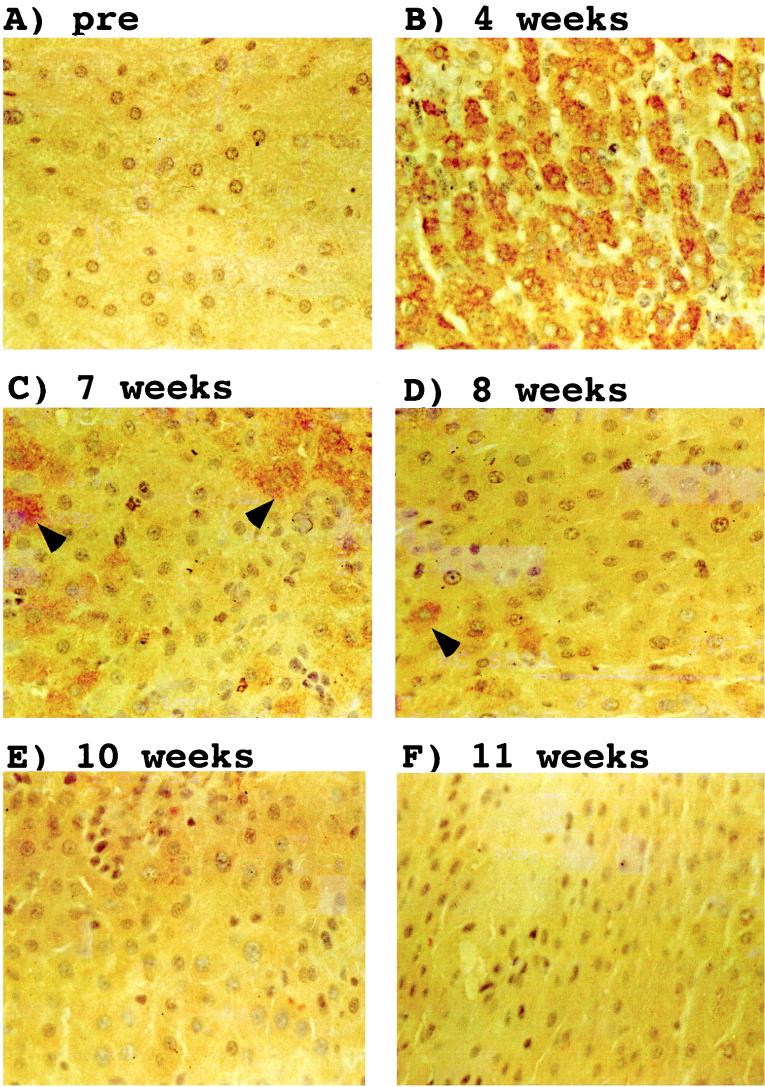
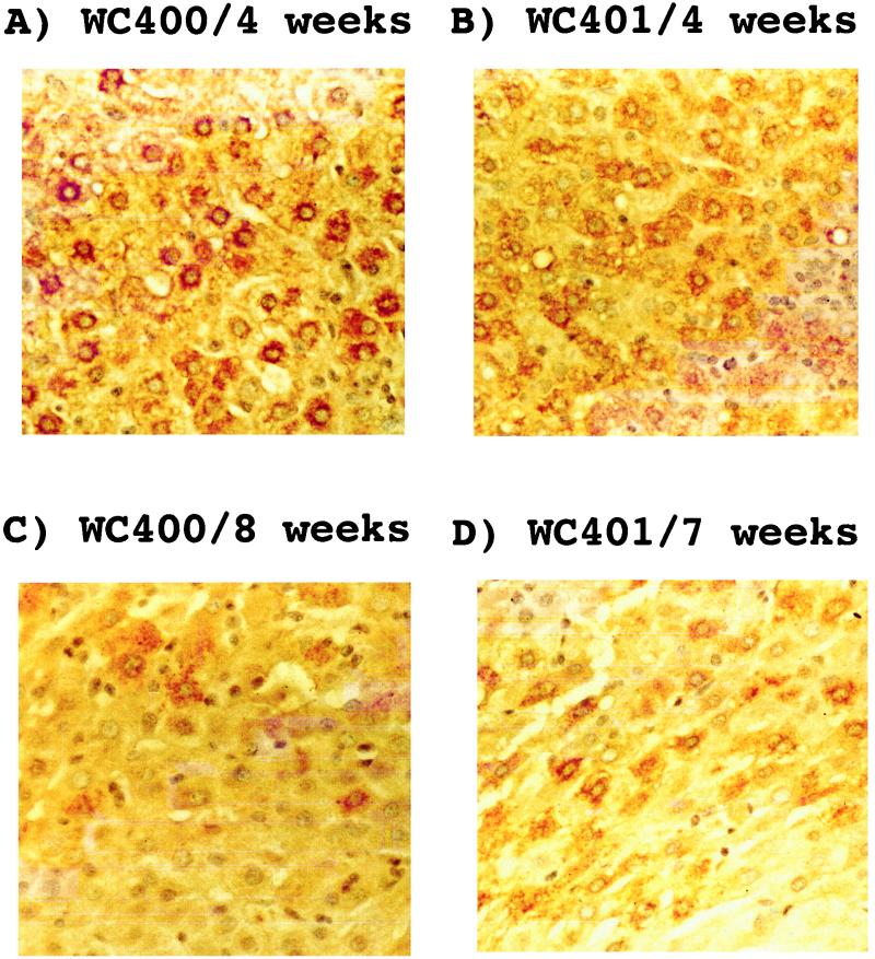
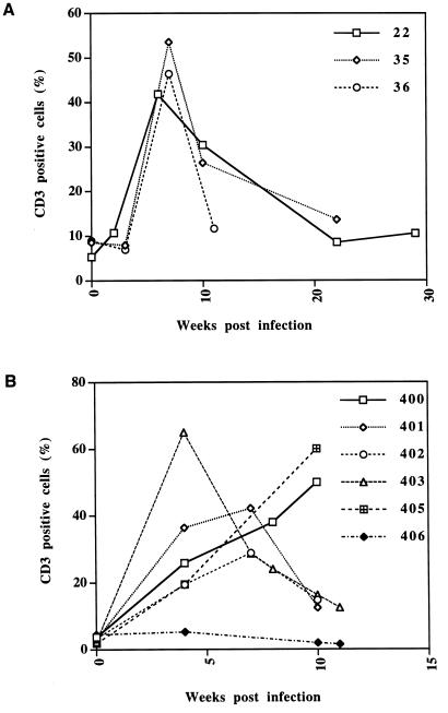
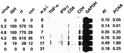
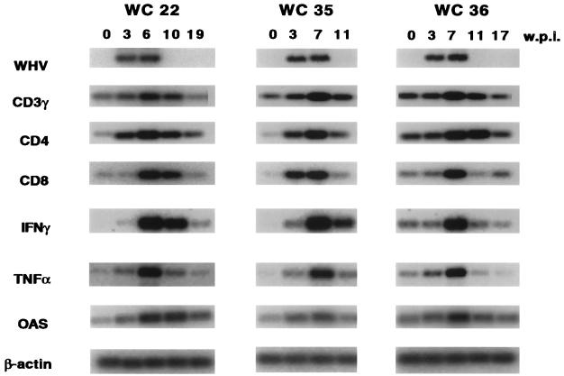
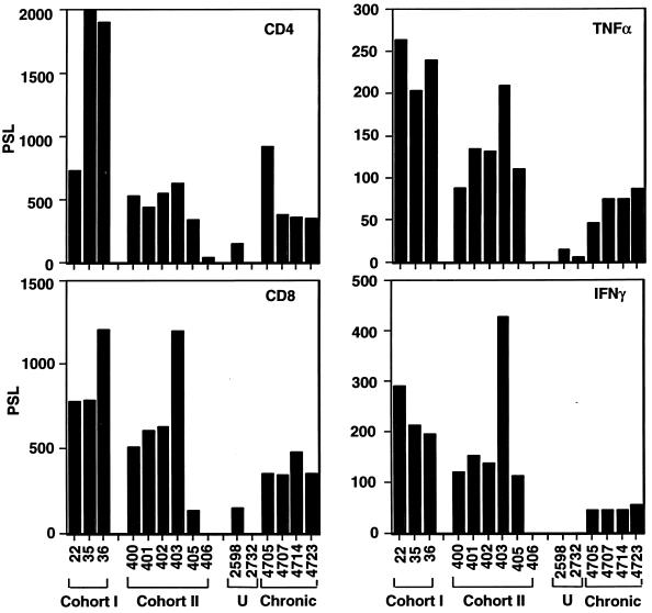
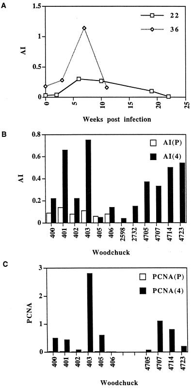
It is well known that hepatitis B virus infections can be transient or chronic, but the basis for this dichotomy is not known. To gain insight into the mechanism responsible for the clearance of hepadnavirus infections, we have performed a molecular and histologic analysis of liver tissues obtained from transiently infected woodchucks during the critical phase of the recovery period. We found as expected that clearance from transient infections occurred subsequent to the appearance of CD4(+) and CD8(+) T cells and the production of interferon gamma and tumor necrosis factor alpha in the infected liver. These events were accompanied by a significant increase in apoptosis and regeneration of hepatocytes. Surprisingly, however, accumulation of virus-free hepatocytes was delayed for several weeks following this initial influx of lymphocytes. In addition, we observed that chronically infected animals can exhibit levels of T-cell accumulation, cytokine expression, and apoptosis that are comparable with those observed during the initial phase of transient infections. Our results are most consistent with a model for recovery predicting replacement of infected hepatocytes with regenerated cells, which by unknown mechanisms remain protected from reinfection in animals that can be cured.


Figures

FIG. 1
FIG. 1
Resolution of the transient WHV infection from the liver of woodchuck 403. Sections from ethanol-acetic acid-fixed liver tissues obtained at the indicated time points were reacted with a rabbit WHV core antigen antibody. The antibody was detected by staining with immunoperoxidase. Hepatocytes that express core antigen are marked with arrows. Magnification, ×388. pre, preinfection.
FIG. 2
FIG. 2
Piecemeal recovery from transient WHV infections in woodchucks 400 and 401. Liver sections were treated and processed as described in the legend to Fig. 1. Magnification, ×400.
FIG. 3
FIG. 3
Infiltration of CD3+ T cells into the livers of WHV-infected woodchucks. The graphs indicate the number of CD3+ cells, not including those in the portal tract, per hepatocyte during the transient infections in woodchucks belonging to cohort I (A) and cohort II (B).
FIG. 4
FIG. 4
Transient WHV infection in woodchuck 38. The left side of the figure shows the results from the analysis of WHV DNA markers during transient WHV infection as determined previously by Kajino et al. (14). The types of values shown in the four leftmost columns are as follows: virus, the number of virions per milliliter of serum (multiplied by 10−9); ISH, the fraction (percentage) of hepatocytes that showed a positive signal in an in situ hybridization (ISH) assay with a WHV probe; and rf and ccc, the copy numbers of replicative form (rf) and cccDNA, respectively, relative to the total number of hepatocytes. The central panel shows the results from the RNase protection analysis with RNA obtained at the indicated number of weeks p.i. (w.p.i.). The right side shows the results from the histological examination of liver sections for apoptotic hepatocytes and for hepatocytes with nuclear PCNA staining. The columns contain the following types of values: the AI, the fraction of hepatocytes that showed morphological signs of apoptosis, and the fraction of hepatocytes with nuclei that were positive for PCNA, respectively.
FIG. 5
FIG. 5
Analysis of WHV and cellular mRNAs in transiently infected woodchucks by RT-PCR. The results of the RT-PCR analysis to detect mRNAs corresponding to WHV, CD3-γ, CD4, and CD8-α and the cytokines IFN-γ and TNF-α and for 2′-5′ OAS and β-actin (details of the procedures are described in Materials and Methods) are shown. w.p.i., weeks p.i.; WC, woodchuck.
FIG. 6
FIG. 6
Analysis of cellular mRNAs in transiently and chronically infected woodchucks by RNase protection. The histograms summarize the results obtained from RNase protection experiments with woodchucks in cohorts I and II as well as two uninfected (U) woodchucks (2598 and 2732) and four chronically infected animals (4705, 4707, 4714, and 4723) (the probes used are indicated in the upper right corner of each histogram). Quantitation of the RNase protection analysis was performed with a phosphorimager. The values were expressed in arbitrary units (PSL).
FIG. 7
FIG. 7
Determination of the AI and expression of PCNA in transiently and chronically infected woodchucks. (A) The results of determinations of apoptotic cells counts from slides of tissue from woodchucks 22 and 36 at the indicated time points. (B) The results of determinations of apoptotic cell counts of cohort II animals prior to infection (P) and 4 weeks p.i. (4). Panel B also shows the results obtained with two uninfected control animals, 2598 and 2732, and four chronically infected woodchucks, 4705, 4707, 4714, and 4723. The AI is the fraction (percentage) of apoptotic hepatocytes per normal hepatocyte. (C) The results of the analysis of PCNA-positive hepatocytes of sections from cohort II animals and the four chronically infected woodchucks.

References

    1. Bursch W, Paffe S, Putz B, Barthel G, Schulte-Hermann R. Determination of the length of the histological stages of apoptosis in normal liver and in altered hepatic foci of rats. Carcinogenesis. 1990;11:847–853. - PubMed
    1. Chirmule N, Moscioni A D, Qian Y, Qian R, Chen Y, Wilson J M. Fas-Fas ligand interactions play a major role in effector functions of cytotoxic T lymphocytes after adenovirus vector-mediated gene transfer. Hum Gene Ther. 1999;10:259–269. - PubMed
    1. Columbano A, Ledda-Columbano G M, Coni P P, Faa G, Liguori C, Santa Cruz G, Pani P. Occurrence of cell death (apoptosis) during the involution of liver hyperplasia. Lab Investig. 1985;52:670–675. - PubMed
    1. Cote P J, Korba B E, Steinberg H, Ramirez M C, Baldwin B, Hornbuckle W E, Tennant B C, Gerin J L. Cyclosporin A modulates the course of woodchuck hepatitis virus infection and induces chronicity. J Immunol. 1991;146:3138–3144. - PubMed
    1. Dienes H P, Purcell R H, Popper H, Ponzetto A. The significance of infections with two types of viral hepatitis demonstrated by histologic features in chimpanzees. J Hepatol. 1990;10:77–84. - PubMed

Publication types

MeSH terms