. 2000 Feb;74(3):1506-12.
doi: 10.1128/jvi.74.3.1506-1512.2000.

Two patches of amino acids on the E2 DNA binding domain define the surface for interaction with E1

Affiliations

Two patches of amino acids on the E2 DNA binding domain define the surface for interaction with E1

G Chen et al. J Virol. 2000 Feb.

Abstract

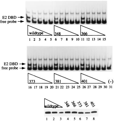
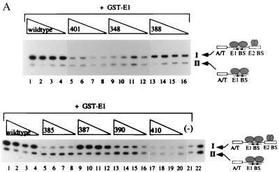
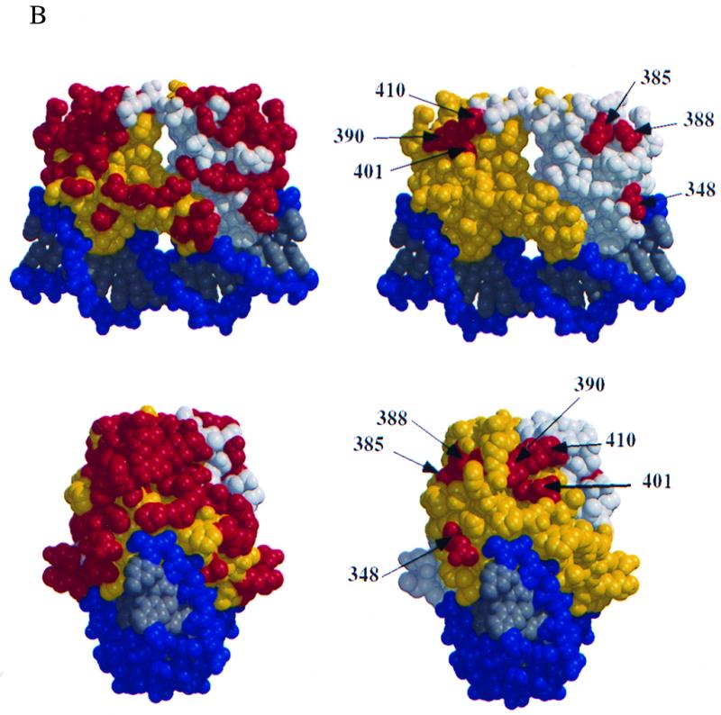
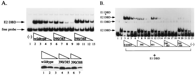
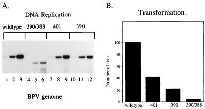
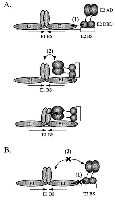
The E1 and E2 proteins from bovine papillomavirus bind cooperatively to the viral origin of DNA replication (ori), forming a complex which is essential for initiation of DNA replication. Cooperative binding has two components, in which (i) the DNA binding domains (DBDs) of the two proteins interact with each other and (ii) the E2 transactivation domain interacts with the helicase domain of E1. By generating specific point mutations in the DBD of E2, we have defined two patches of amino acids that are involved in the interaction with the E1 DBD. These same mutations, when introduced into the viral genome, result in severely reduced replication of the viral genome, as well as failure to transform mouse cells in tissue culture. Thus, the interaction between the E1 and E2 DBDs is important for the establishment of the viral genome as an episome and most likely contributes to the formation of a preinitiation complex on the viral ori.


Figures

FIG. 1
FIG. 1
DNA binding activities of E2 DBD point mutant proteins. Levels of mutant E2 DBDs were determined by Western blot analysis using a polyclonal antibody to E2 and quantitated by using an IS 1000 Digital Imaging system. Three different dilutions of crude E. coli extracts containing the wt E2 DBD (lanes 1 to 3, bottom panel) were used as standards for levels of expression of five different mutant proteins (lanes 4 to 8, bottom panel), and equivalent amounts of protein were used to test for DNA binding activity by EMSA. Twofold titrations of the wt protein (lanes 1 to 5, top panel) or mutant proteins (lanes 6 to 30, top panel) were analyzed on a 5% polyacrylamide gel. Lane 31 contained probe alone.
FIG. 2
FIG. 2
(A) Six mutant proteins are defective for cooperative binding with E1. Two different probes, one containing the BPV minimal ori with a high-affinity E2 binding site (I) and one containing an E1 binding site alone (II), were incubated with 6 ng of GST-E1 alone (lane 21, bottom) or with either the wt E2 DBD (lanes 1 to 4, top and bottom), or seven mutant E2 DBDs (lanes 5 to 16, top, and lanes 5 to 20, bottom). Equal quantities of wt and mutant proteins, based on Western blot analysis, were used in four twofold titrations. Probes bound by GST-E1 were recovered with glutathione-agarose beads and analyzed on a 6% urea gel. E2-dependent stimulation of binding was measured by comparing the amounts of probes II and I recovered. (B) Five mutations which do not affect DNA binding form two distinct patches on the E2 DBD. Shown are two different views of a space-filling model of the E2 DBD bound to its cognate binding site, created by the BOBSCRIPT program and Raster3D (6, 10, 17). In the images on the right, all mutations which were made on the surface of the E2 DBD are in red. The images on the left show the same two views with mutations that affect the ability of the E2 DBD to interact with E1 in red.
FIG. 2
FIG. 2
(A) Six mutant proteins are defective for cooperative binding with E1. Two different probes, one containing the BPV minimal ori with a high-affinity E2 binding site (I) and one containing an E1 binding site alone (II), were incubated with 6 ng of GST-E1 alone (lane 21, bottom) or with either the wt E2 DBD (lanes 1 to 4, top and bottom), or seven mutant E2 DBDs (lanes 5 to 16, top, and lanes 5 to 20, bottom). Equal quantities of wt and mutant proteins, based on Western blot analysis, were used in four twofold titrations. Probes bound by GST-E1 were recovered with glutathione-agarose beads and analyzed on a 6% urea gel. E2-dependent stimulation of binding was measured by comparing the amounts of probes II and I recovered. (B) Five mutations which do not affect DNA binding form two distinct patches on the E2 DBD. Shown are two different views of a space-filling model of the E2 DBD bound to its cognate binding site, created by the BOBSCRIPT program and Raster3D (6, 10, 17). In the images on the right, all mutations which were made on the surface of the E2 DBD are in red. The images on the left show the same two views with mutations that affect the ability of the E2 DBD to interact with E1 in red.
FIG. 3
FIG. 3
(A) DNA binding activities of double mutant E2 DBDs. The double mutant proteins 390/385 (lanes 4 and 5, bottom) and 390/388 (lanes 6 and 7, bottom) were quantitated with wt E2 DBD standards (lanes 1 to 3, bottom) as described in the legend to Fig. 1. Equal quantities of mutant and wt E2 DBDs were then tested for DNA binding activity by EMSA using four twofold dilutions of wt E2 DBD (lanes 2 to 5, top), 390/385 (lanes 6 to 9, top), or 390/388 (lanes 10 to 13, top). (B) The double mutant proteins 390/385 and 390/388 failed to interact with E1. To determine the abilities of the mutant proteins to interact with the E1 DBD, an EMSA was performed with a probe containing the BPV minimal ori with a high-affinity E2 binding site. A 0.4-ng sample of the E1 DBD (aa 142 to 308) was incubated with four different twofold titrations of E. coli extracts containing the wt E2 DBD (lanes 3 to 6) or the mutant protein 390 (lanes 7 to 10), 401 (lanes 11 to 14), 390/385 (lanes 15 to 18), or 390/388 (lanes 19 to 22). Lane 1 contained 0.4 ng of the E1 DBD (aa 142 to 308) alone, lane 2 contained the wt E2 DBD alone, and lane 23 contained probe alone.
FIG. 4
FIG. 4
(A) The mutations in the E2 DBD affect replication of the viral genome. The BPV genome was mutated at position(s) 390, 401, or 390 and 388. Transient replication assays were performed with mouse C127 cells, and low-molecular-weight DNA was digested with the restriction enzymes DpnI and HindIII. Replication of the wt (lanes 1 to 3) or the 390/388 (lanes 4 to 6), 401 (lanes 7 to 9), or 390 (lanes 10 to 12) mutant genome was measured at 24, 48, and 72 h posttransfection. In lanes 4 to 6, the viral DNA containing mutations at positions 390 and 388 shows increased mobility due to the presence of an additional HindIII site generated by the mutation. (B) Mutations which affect the E2 DBD-E1 interaction affect viral transformation. The wt genome and the genome mutated at position(s) 390, 401, or 390 and 388 were transfected into C127 cells. After 2 weeks, plates were stained with methylene blue and transformed foci were counted.
FIG. 5
FIG. 5
Stability of wt and mutant E2 proteins. COS cells were transfected with a pCG expression vector encoding wt E2 or the 390/388 mutant protein. At 20 h after transfection, protein synthesis was blocked by the addition of 25 μg cycloheximide per ml. Cells were harvested, and the levels of E2 protein after 1 h (lanes 4 and 7) and 2 h (lanes 5 and 8) in the presence of cycloheximide were compared to E2 levels in the absence of cycloheximide (lanes 3 and 6) by Western blotting using a polyclonal antiserum to E2. Lane 1 contained purified E2 protein; lane 2 contained extract from untransfected cells. The calculated half-life of both the wt and the 390/388 mutant protein, based on quantitation of the Western blot, was approximately 50 min.
FIG. 6
FIG. 6
Model of cooperative binding of E1 and E2 to the BPV minimal ori. (A) Cooperative binding between E1 and E2 involves two separate interactions: interaction 1 between the E1 and E2 DBDs and interaction 2 between the E1 helicase domain and the E2 activation domain (AD). As the first required step in the cooperative binding of E1 and E2 on the BPV minimal ori, the E2 DBD interacts with the E1 DBD (interaction 1). This interaction results in bending or kinking of the DNA. As a consequence of the induced DNA bend, the E2 activation domain is placed in a position where it can effectively interact with the E1 helicase domain. The productive interaction between the E2 activation domain and E1 completes the second step in the cooperative binding of E1 and E2 on the ori. (B) Mutations in the E2 DBD which result in failure to interact with the E1 DBD result in loss of the interaction between E1 and E2 despite a wt and functional E2 activation domain.

References

    1. Abroi A, Kurt R, Ustav M. Transcriptional and replicational activation functions in the bovine papillomavirus type 1 E2 protein are encoded by different structural determinants. J Virol. 1996;70:6169–6179. - PMC - PubMed
    1. Benson J D, Howley P M. Amino-terminal domains of the bovine papillomavirus type 1 E1 and E2 proteins participate in complex formation. J Virol. 1995;69:4364–4372. - PMC - PubMed
    1. Berg M, Stenlund A. Functional interactions between papillomavirus E1 and E2 proteins. J Virol. 1997;71:3853–3863. - PMC - PubMed
    1. Chen G, Stenlund A. Characterization of the DNA-binding domain of the bovine papillomavirus replication initiator E1. J Virol. 1998;72:2567–2576. - PMC - PubMed
    1. Chen L, Glover J N M, Hogan P G, Rao A, Harrison S C. Structure of the DNA-binding domains from NFAT, Fos and Jun bound specifically to DNA. Nature. 1998;392:42–48. - PubMed

Publication types

MeSH terms

LinkOut - more resources