. 2000 Feb;74(3):1513-23.
doi: 10.1128/jvi.74.3.1513-1523.2000.

Alpha/beta interferons potentiate virus-induced apoptosis through activation of the FADD/Caspase-8 death signaling pathway

Affiliations

Alpha/beta interferons potentiate virus-induced apoptosis through activation of the FADD/Caspase-8 death signaling pathway

S Balachandran et al. J Virol. 2000 Feb.

Abstract

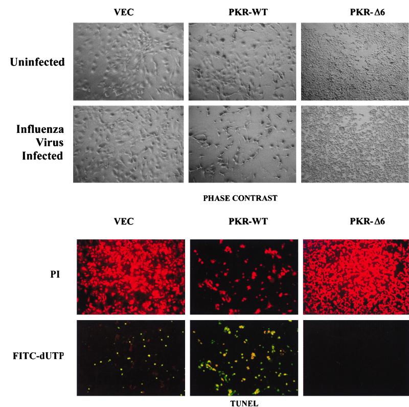
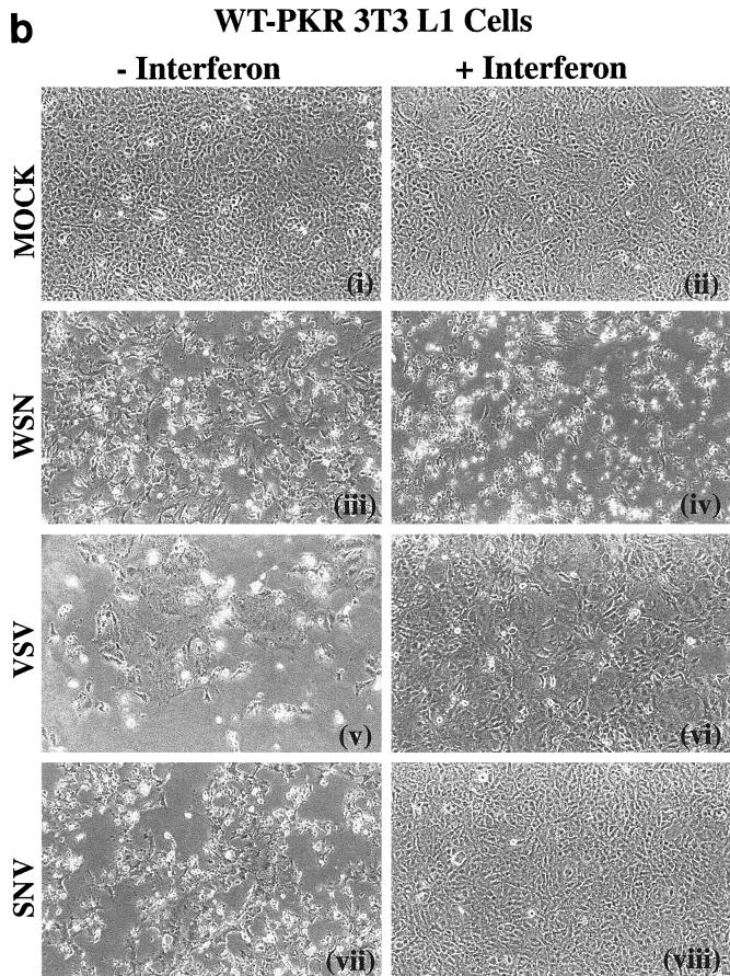
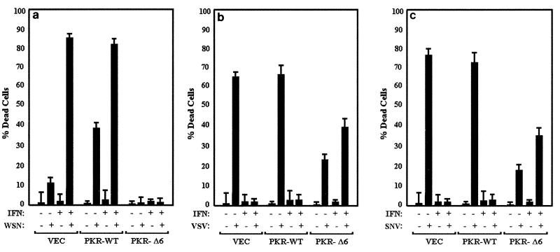
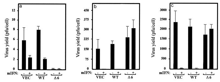
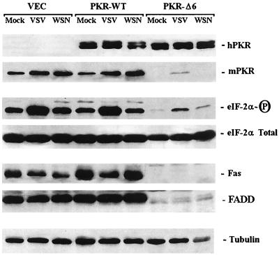
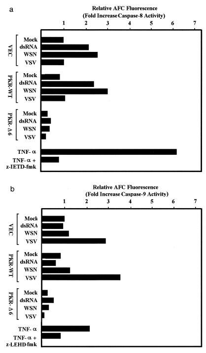
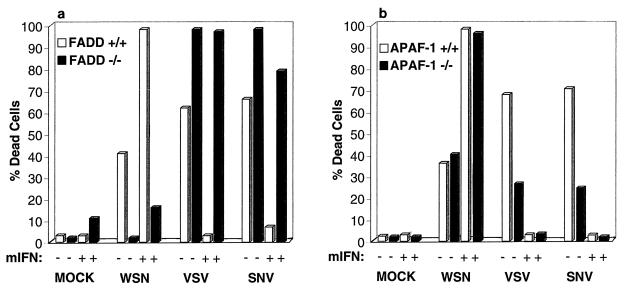
Interferon (IFN) mediates its antiviral effects by inducing a number of responsive genes, including the double-stranded RNA (dsRNA)-dependent protein kinase, PKR. Here we report that inducible overexpression of functional PKR in murine fibroblasts sensitized cells to apoptosis induced by influenza virus, while in contrast, cells expressing a dominant-negative variant of PKR were completely resistant. We determined that the mechanism of influenza virus-induced apoptosis involved death signaling through FADD/caspase-8 activation, while other viruses such as vesicular stomatitis virus (VSV) and Sindbis virus (SNV) did not significantly provoke PKR-mediated apoptosis but did induce cytolysis of fibroblasts via activation of caspase-9. Significantly, treatment with IFN-alpha/beta greatly sensitized the fibroblasts to FADD-dependent apoptosis in response to dsRNA treatment or influenza virus infection but completely protected the cells against VSV and SNV replication in the absence of any cellular destruction. The mechanism by which IFN increases the cells' susceptibility to lysis by dsRNA or certain virus infection is by priming cells to FADD-dependent apoptosis, possibly by regulating the activity of the death-induced signaling complex (DISC). Conversely, IFN is also able to prevent the replication of viruses such as VSV that avoid triggering FADD-mediated DISC activity, by noncytopathic mechanisms, thus preventing destruction of the cell.


Figures

FIG. 1
FIG. 1
Cells inducibly expressing PKR are sensitive to WSN-induced apoptosis. Murine 3T3 L1 cells (VEC, control cells carrying vector alone; PKR-WT, cells overexpressing PKR under control of a tetracycline/doxycycline-inducible promoter; PKR-Δ6, cells overexpressing a dominant-negative variant of PKR under the same inducible promoter) were infected with WSN at an MOI of 10. (Top) Cells mock infected or infected with WSN were photographed 24 h postinfection (magnification, ×100). (Bottom) WSN-infected cells were fixed and incubated with the TUNEL reaction mixture (see Materials and Methods) to detect apoptotic cells and subsequently stained with propidium iodide to detect all cells (magnification, ×100).
FIG. 2
FIG. 2
Micrographs of WSN-, VSV-, and SNV-infected cells inducibly overexpressing PKR and treated with IFN. 3T3 L1 cells carrying vector alone (a) or PKR-overexpressing 3T3 L1 cells (b) were mock infected or infected with WSN, VSV, or SNV at an MOI of 10 in the presence (+) or absence (−) of murine fibroblast IFN-α/β (1,000 U/ml) pretreatment. Photomicrographs were taken 24 (WSN) or 48 (VSV and SNV) h postinfection at a magnification of ×100. FIG. 2—Continued.
FIG. 2
FIG. 2
Micrographs of WSN-, VSV-, and SNV-infected cells inducibly overexpressing PKR and treated with IFN. 3T3 L1 cells carrying vector alone (a) or PKR-overexpressing 3T3 L1 cells (b) were mock infected or infected with WSN, VSV, or SNV at an MOI of 10 in the presence (+) or absence (−) of murine fibroblast IFN-α/β (1,000 U/ml) pretreatment. Photomicrographs were taken 24 (WSN) or 48 (VSV and SNV) h postinfection at a magnification of ×100. FIG. 2—Continued.
FIG. 3
FIG. 3
IFN sensitizes infected cells to or protects them from apoptosis. IFN-treated or untreated 3T3 L1 control cells (VEC) or cells inducibly overexpressing wild-type PKR (PKR-WT) or a dominant-negative variant (PKR-Δ6) were mock infected or infected with WSN (a), VSV (b), or SNV (c) at an MOI of 10 in the presence or absence of IFN pretreatment; 24 (WSN) or 48 (VSV and SNV) h later, trypan blue exclusion analysis was performed. Data represent the mean from three experiments ± standard deviation.
FIG. 4
FIG. 4
Effects of PKR and IFN on viral production. IFN-treated or untreated 3T3 L1 cells (VEC) inducibly expressing wild-type PKR (WT) or dominant-negative variant PKR-Δ6 (Δ6) were infected with WSN (a), VSV (b), or SNV (c) at an MOI of 10; 24 (WSN) or 48 (VSV and SNV) h later, samples of medium from the infected cells were retrieved and viral production was determined by plaque assay. Data represent means of three experiments ± standard deviation.
FIG. 5
FIG. 5
Protein analysis of PKR, eIF2α, and Fas. Murine 3T3 L1 cells (VEC) inducibly expressing wild-type PKR (PKR-WT) or a dominant-negative variant (PKR-Δ6) were infected with VSV or WSN; 24 h postinfection, equal amounts of cell lysates were examined by Western blotting using antibodies to human PKR (hPKR), the endogenous murine PKR (mPKR), phosphorylated eIF2α (eIF-2α-P) total eIF2α, Fas, and FADD. Similar levels of tubulin in each lane confirm that equivalent amounts of protein were loaded.
FIG. 6
FIG. 6
Caspase activity in cells infected with WSN or VSV. Control 3T3 L1 cells (VEC) or 3T3 L1 cells overexpressing wild-type PKR (PKR-WT) or a dominant-negative variant (PKRΔ6) were transfected with poly(I-C) by using Lipofectamine (Gibco-BRL) or infected with WSN or VSV at an MOI of 10. After 24 h, cell lysates were prepared and incubated with either z-IETD-AFC, the caspase-8 fluorogenic substrate (a), or z-LEHD-AFC, the caspase-9 fluorogenic substrate (Apoalert; Clontech) (b). Samples were excited at 400 nm, and fluorescent emissions were read at 505 nm on a fluorometer. As controls, cell lysates prepared from 3T3 L1 cells treated for 18 h with 50 ng of murine TNF-α (R&D Systems) per ml were preincubated with or without the caspase-8-specific inhibitor z-LEHD-fmk or the caspase-9 specific blocker z-LEHD-fmk prior to incubation with the fluorogenic substrates as described above. Data represent means of two experiments.
FIG. 7
FIG. 7
Murine fibroblasts lacking FADD but not Apaf-1 show resistance to WSN-induced apoptosis. Fibroblasts lacking FADD (a) or Apaf-1 (b) were infected with WSN, VSV, or SNV at an MOI of 10; 24 (WSN) or 48 (VSV and SNV) h postinfection, cell viability was determined by trypan blue exclusion analysis. Data represent means of two experiments.

References

    1. Abraham N, Stojl D F, Duncan P I, Methot N, Ishii T, Dube M, Vanderhyden B C, Atkins H L, Gray D A, McBurney M W, Koromilas A E, Brown E G, Sonenberg N, Bell J C. Characterization of transgenic mice with a targeted disruption of the catalytic domain of the double-stranded RNA-dependent protein kinase, PKR. J Biol Chem. 1999;247:5953–5962. - PubMed
    1. Balachandran S, Kim C N, Yeh W-C, Mak T W, Bhalla K N, Barber G N. Activation of the dsRNA-dependent protein kinase, PKR, induces apoptosis through FADD-mediated death signaling. EMBO J. 1998;17:6888–6902. - PMC - PubMed
    1. Banerjee A K. Transcription and replication of rhabdoviruses. Microbiol Rev. 1987;51:66–87. - PMC - PubMed
    1. Barber G N, Wambach M, Wong M L, Dever T E, Hinnebusch A G, Katze M G. Translational regulation by the interferon-induced double-stranded RNA-activated 68-kDa protein kinase. Proc Natl Acad Sci USA. 1993;90:4621–4625. - PMC - PubMed
    1. Chinnaiyan A M, O'Rouke K, Tewari M, Dixit V M. FADD, a novel death domain-containing protein, interacts with the death domain of Fas and initiates apoptosis. Cell. 1995;81:505–512. - PubMed

Publication types

MeSH terms

LinkOut - more resources