. 2000 Jan 1;20(1):43-50.
doi: 10.1523/JNEUROSCI.20-01-00043.2000.

Rescue of hearing, auditory hair cells, and neurons by CEP-1347/KT7515, an inhibitor of c-Jun N-terminal kinase activation

Affiliations

Rescue of hearing, auditory hair cells, and neurons by CEP-1347/KT7515, an inhibitor of c-Jun N-terminal kinase activation

U Pirvola et al. J Neurosci. .

Abstract

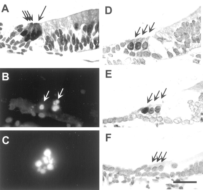
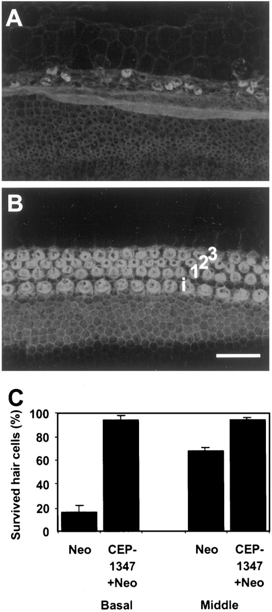
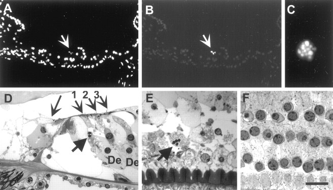
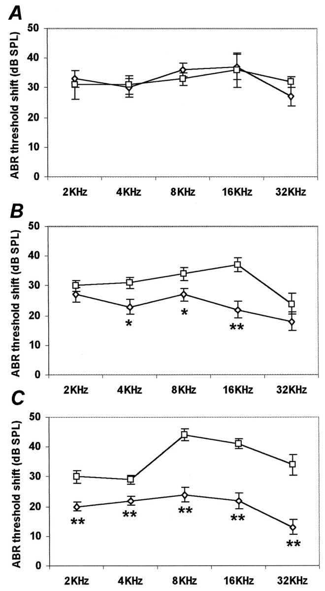
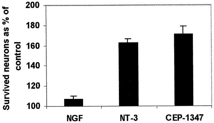
We have studied the mechanisms of auditory hair cell death after insults in vitro and in vivo. We show DNA fragmentation of hair cell nuclei after ototoxic drug and intense noise trauma. By using phospho-specific c-Jun-N-terminal kinase (JNK) and c-Jun antibodies in immunohistochemistry, we show that the JNK pathway, associated with stress, injury, and apoptosis, is activated in hair cells after trauma. CEP-1347, a derivative of the indolocarbazole K252a, is a small molecule that has been shown to attenuate neurodegeneration by blocking the activation of JNK (). Subcutaneously delivered CEP-1347 attenuated noise-induced hearing loss. The protective effect was demonstrated by functional tests, which showed less hearing threshold shift in CEP-1347-treated than in nontreated guinea pigs, and by morphometric methods showing less hair cell death in CEP-1347-treated cochleas. In organotypic cochlear cultures, CEP-1347 prevented neomycin-induced hair cell death. In addition to hair cells, CEP-1347 promoted survival of dissociated cochlear neurons. These results suggest that therapeutic intervention in the JNK signaling cascade, possibly by using CEP-1347, may offer opportunities to treat inner ear injuries.


Figures

Fig. 1.
Fig. 1.
Hair cell death and JNK and c-Jun phosphorylation in neomycin-exposed (100 μm) cochlear explants of neonatal rats. The specimens were embedded in paraffin and cut in transverse (midmodiolar) plane. A, One row of calbindin-immunoreactive inner hair cells and three rows of outer hair cells (arrows) are seen in nonexposed explants. B, TUNEL-stained outer hair cell nuclei (arrows) are seen in cultures exposed to neomycin for 12 hr. C, Higher magnification of an outer hair cell nucleus showing TUNEL-positive DNA fragmentation. D, E, Phospho-JNK and phospho-c-Jun immunolabeling, respectively, are found in the nuclei of hair cells (arrows) exposed to neomycin for 6 hr. F, Phospho-c-Jun-immunoreactive hair cells are not seen in cultures coincubated with neomycin and CEP-1347 (500 nm) for 6 hr. Arrows point to hair cells. Scale bar: A,B, D–F, 27 μm; C, 10 μm.
Fig. 2.
Fig. 2.
CEP-1347 (500 nm) prevents hair cell loss in cochlear explants of neonatal rats exposed to 100 μm neomycin for 48 hr. A, Neomycin causes severe hair cell degeneration in the basal turn of the cochlea, as shown in phalloidin-labeled confocal images. B, CEP-1347 prevents hair cell loss in the basal turn of the cochlea.i, Inner hair cell; 1, 2, 3, rows of outer hair cells. C, Quantification of numbers of survived outer hair cells in basal and middle turns. Histograms and bars represent mean ± SEM for three experiments, each including four explants of both the control and CEP-1347 group. Scale bar:A,B, 50 μm.
Fig. 3.
Fig. 3.
Hair cell death in the guinea pig cochlea 1 d after intense noise exposure (120 dB, 4 kHz, 6 hr). A,B, The transverse (midmodiolar) paraffin section of the organ of Corti in the second cochlear turn is doublestained with DAPI nuclear stain (A) and with TUNEL method (B). The three outer hair cells (arrow) show distorted nuclei and are TUNEL-positive. Supporting cells, seen below the hair cells, are not TUNEL-labeled.C, At high magnification, a TUNEL-labeled paraffin section shows an outer hair cell nucleus with DNA fragmentation.D, A toluidine blue-stained, resin-embedded semithin section of the organ of Corti in transverse plane shows an inner hair cell (large arrow) and three rows of OHCs (small arrows). Only OHC1 shows a fragmented nucleus (thick arrow). The section is from the area of scattered hair cell loss. De, Deiter's cells. E, A toluidine blue-stained, resin-embedded semithin section in horizontal plane and at the level of hair cell nuclei. The section is from the region of maximal trauma. Most hair cells are lost, except one outer hair cell that shows a fragmented nucleus (thick arrow).F, A section, prepared as in E, from nontraumatized region of the organ of Corti. Outer hair cells of the three rows are present, and their nuclei are not fragmented. Scale bar:A,B, 80 μm; C, 10 μm;D, 18 μm; E,F, 15 μm.
Fig. 4.
Fig. 4.
CEP-1347 attenuates intense noise-induced hearing loss in vivo. Hearing threshold shifts were assessed by auditory brainstem testing 2 (A), 6 (B), and 14 (C) d after exposing control (■; n = 4 at 2 d, 8 at 6 d, 17 at 14 d) and CEP-1347-treated (⋄;n = 5 at 2 d, 11 at 6 d, 17 at 14 d) guinea pigs to noise (120 dB, 4 kHz, 6 hr). Results show mean ± SEM. Six and 14 d after exposure, the average threshold shifts of control animals are significantly greater than that of CEP-1347-treated animals. **p < 0.01; *p < 0.05; Student's t test.
Fig. 5.
Fig. 5.
CEP-1347 attenuates intense noise-induced hair cell loss in vivo. Quantification of hair cell numbers in vehicle-treated, noise-exposed control (A;n = 13) and CEP-1347-treated guinea pig cochleas (B;n = 13) 2 weeks after noise exposure (120 dB, 4 kHz, 6 hr). In the averaged cytocochleograms, the percentages of IHCs and OHCs are plotted for the entire length of the organ of Corti. Maximal damage is located in a region ∼50% from the round window. CEP-1347 treatment attenuates hair cell loss.
Fig. 6.
Fig. 6.
Effects of CEP-1347 (500 nm), neurotrophin-3 (2 ng/ml), and nerve growth factor (2 ng/ml) on the survival of neonatal cochlear neurons in vitro. Numbers of neurons in cultures with added compounds are expressed as percentage of the number of neurons in control cultures. Neurons were counted after 48 hr in culture. Values represent mean ± SEM from three separate experiments.

References

    1. Basile AS, Huang J-M, Xie C, Webster D, Berlin C, Skolnik P. N-methyl-d-aspartate antagonists limit aminoglycoside antibiotic-induced hearing loss. Nat Med. 1996;2:1338–1343. - PubMed
    1. Behrens A, Sibilia M, Wagner E. Amino-terminal phosphorylation of c-Jun regulates stress-induced apoptosis and cellular proliferation. Nat Genet. 1999;21:326–329. - PubMed
    1. Borasio GD, Horstmann S, Anneser JMH, Neff NT, Glicksman MA. CEP-1347/KT7515, a JNK pathway inhibitor, supports the in vitro survival of chick embryonic neurons. NeuroReport. 1998;9:1435–1439. - PubMed
    1. Brady R, Zaidi SIA, Mayer C, Katz DM. BDNF is a target-derived survival factor for arterial baroreceptor and chemoafferent primary sensory neurons. J Neurosci. 1999;19:2131–2142. - PMC - PubMed
    1. Clerici WJ, Hensley K, DiMartino DL, Butterfield DA. Direct detection of ototoxicant-induced reactive oxygen species generation in cochlear explants. Hear Res. 1996;98:116–124. - PubMed

Publication types

MeSH terms