. 2000 Jan 1;20(1):66-75.
doi: 10.1523/JNEUROSCI.20-01-00066.2000.

Nicotinic receptor activation in human cerebral cortical interneurons: a mechanism for inhibition and disinhibition of neuronal networks

Affiliations

Nicotinic receptor activation in human cerebral cortical interneurons: a mechanism for inhibition and disinhibition of neuronal networks

M Alkondon et al. J Neurosci. .

Abstract

Cholinergic control of the activity of human cerebral cortical circuits has long been thought to be accounted for by the interaction of acetylcholine (ACh) with muscarinic receptors. Here we report the discovery of functional nicotinic receptors (nAChRs) in interneurons of the human cerebral cortex and discuss the physiological and clinical implications of these findings. The whole-cell mode of the patch-clamp technique was used to record responses triggered by U-tube application of the nonselective agonist ACh and of the alpha7-nAChR-selective agonist choline to interneurons visualized by means of infrared-assisted videomicroscopy in slices of the human cerebral cortex. Choline induced rapidly desensitizing whole-cell currents that, being sensitive to blockade by methyllycaconitine (MLA; 50 nM), were most likely subserved by an alpha7-like nAChR. In contrast, ACh evoked slowly decaying whole-cell currents that, being sensitive to blockade by dihydro-beta-erythroidine (DHbetaE; 10 microM), were most likely subserved by an alpha4beta2-like nAChR. Application of ACh (but not choline) to the slices also triggered GABAergic postsynaptic currents (PSCs). Evidence is provided that ACh-evoked PSCs are the result of activation of alpha4beta2-like nAChRs present in preterminal axon segments and/or in presynaptic terminals of interneurons. Thus, nAChRs can relay inhibitory and/or disinhibitory signals to pyramidal neurons and thereby modulate the activity of neuronal circuits in the human cerebral cortex. These mechanisms, which appear to be retained across species, can account for the involvement of nAChRs in cognitive functions and in certain neuropathological conditions.


Figures

Fig. 1.
Fig. 1.
Photomicrographs of neurons in human cerebral cortical slices. A, B,Typical examples of infrared-assisted videomicroscopic images of a pyramidal neuron (A) and a nonpyramidal neuron/interneuron (B) in human cerebral cortical slices. C, Image of a biocytin-filled interneuron visualized in a formaldehyde-fixed human cerebral cortical slice.D, Neurolucida drawing of the biocytin-filled neuron shown in C.
Fig. 2.
Fig. 2.
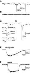
Whole-cell recordings from visually identified interneurons in human cortical slices. A, Samples of spontaneous PSCs and FCTs recorded at −62 mV from a cerebral cortical interneuron. B, Sample recording obtained from the same interneuron after 5 min exposure to bicuculline (10 μm).C, Individual PSCs are shown on an expanded scale.D, An FCT superimposed on a PSC is shown in expanded scale. E, Sample recording obtained from a neuron during a 6 sec U-tube application of quisqualate (20 μm) at −68 mV. F, Sample recording obtained from a neuron during a 6 sec U-tube application of GABA (20 μm) at −62 mV.Solid bars at the top of the traces indicate duration of the agonist pulses. The Cl, F-containing internal solution (see Materials and Methods) was used in most of the experiments (A–D,F), and the methanesulfonate-containing solution (see Materials and Methods) was used in the experiment shown inE.
Fig. 3.
Fig. 3.
Nicotinic currents evoked by agonists applied to interneurons in human cortical slices. Traces, Samples of nicotinic currents evoked by choline (10 mm) in the absence and in the presence of the α7 nAChR-selective antagonist MLA (50 nm) and samples of nicotinic currents evoked by ACh (1 mm) in the absence and in the presence of the α4β2 nAChR-selective antagonist DHβE (10 μm). Samples are from two different neurons. Choline-induced current decayed to the baseline level during the application of the agonist, whereas ACh-induced current maintained a steady-state level in the presence of the agonist. Choline failed to induce any current when the slice was superfused for 5 min with MLA (50 nm)-containing ACSF. ACh-induced slowly decaying current was blocked when the slice was superfused for 5 min with DHβE (10 μm)-containing ACSF. The methanesulfonate-containing internal solution was used to fill the patch pipette. Atropine (1 μm) was present in all recording solutions bathing the slice and in the agonist solutions. Membrane potential, −44 mV.Graph, Average current amplitude evoked by choline in the absence (control) and in the presence of MLA (n= 5 neurons), and average current amplitude elicited by ACh in the absence (control) and in the presence of DHβE (n= 3 neurons). Results are mean ± SE.
Fig. 4.
Fig. 4.
Characteristics of ACh-induced PSCs in human cerebral cortical interneurons. A, Samples of ACh-induced PSCs recorded at different membrane potentials. U-tube application of ACh (1 mm; A, top trace) induced a large inward current associated with isolated inward-going PSCs at −68 mV. The rising phase of the current sampled at a higher frequency (10 kHz instead of 1 kHz) is illustrated by the trace marked with an asterisk (the faster time scale is applicable only to this trace). Note that this rising phase is composed of multiple peaks and is the result of summation of several PSCs. At the null potential (−44 mV; middle trace) for GABA-mediated currents under the present ionic conditions, ACh produced a small inward current, probably representing the postsynaptic nicotinic response of this cell. At 0 mV (bottom trace), ACh induced a burst of outward going PSCs that also showed summation in the beginning of the agonist pulse. The methanesulfonate-containing internal solution (see Materials and Methods) was used in all the experiments illustrated in this figure. B, Samples of ACh-induced outward PSCs recorded at 0 mV under control condition (top trace), 5 min after exposure of the slices to DHβE (10 μm; middle), and 8 min after exposure of the slices to MLA (50 nm;bottom). C, Samples of ACh-evoked PSCs recorded before and 5 min after bath application of TTX (200 nm). D, Samples of ACh-induced PSCs recorded before and 5 min after bath application of bicuculline (10 μm).
Fig. 5.
Fig. 5.
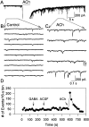
Specificity of ACh effect and time course of ACh-induced PSCs. A, Sample of recordings obtained from a human cortical interneuron exposed for 6 sec to ACh at −62 mV. The ACh-induced, summated PSCs are shown expanded and truncated so that the isolated PSCs that remained well after the agonist pulse can be seen.B, Control PSCs from a small segment of the recording shown on an expanded scale. C, ACh-induced PSCs shown on an expanded scale. D, Summary of the changes in the frequency of PSCs with time caused by a 6 sec, U-tube application of various agents. The patch pipette was filled with the Cl, F-containing internal solution.
Fig. 6.
Fig. 6.
Analysis of ACh-induced PSCs. A,Distribution of peak amplitudes of PSCs before (184 events;left) and after a 6 sec U-tube application of ACh (1 mm, 348 events; right) to a human cerebral cortical interneuron. PSCs were sampled at 5 kHz. Thus, multi-peak events appeared as single-peak events in this analysis. The Gaussian fit of the histogram is shown as a solid line. Membrane potential, 0 mV. B, Cumulative probability plots of peak amplitude histograms and of interevent intervals before and after a 6 sec exposure of the interneuron to ACh (1 mm). Data are from the same experiment as in A. C,Examples of PSCs that show more than one peak (indicated bynumbers at the top) either during the rising phase or during the decay phase of the currents. Traces were sampled at 20 kHz in order to reveal multiple peaks.
Fig. 7.
Fig. 7.
ACh-induced PSCs in acutely dissociated human cerebral cortical neurons. A, Image of a neuron acutely dissociated by mechanical means (Barbosa et al., 1996) from a human cerebral cortical slice. B, Sample recordings obtained from a human cerebral cortical neuron exposed to ACh before (B1) and after (B2) its perfusion with TTX (200 nm)-containing external solution.C, Sample recording obtained from a neuron that showed an inward nicotinic current in response to ACh (1 mm) in the presence of TTX. Membrane potential = −62 mV. The Cl, F-containing internal solution was used to fill the recording pipette. The bathing and the agonist-containing solutions had atropine (1 μm).
Fig. 8.
Fig. 8.
Proposed scheme of the involvement of nAChRs in the control of the overall activity of a hypothetical neuronal circuit in the human cerebral cortex. Sites a, b, andc are presumed to contain nAChRs.

References

    1. Albuquerque EX, Alkondon M, Pereira EFR, Castro NG, Schrattenholz A, Barbosa CTF, Bonfante-Cabarcas R, Aracava Y, Eisenberg H, Maelicke A. Properties of neuronal nicotinic acetylcholine receptors: pharmacological characterization and modulation of synaptic function. J Pharmacol Exp Ther. 1996;280:1117–1136. - PubMed
    1. Alkondon M, Pereira EFR, Barbosa CTF, Albuquerque EX. Neuronal nicotinic acetylcholine receptor activation modulates γ-aminobutyric acid release from CA1 neurons of rat hippocampal slices. J Pharmacol Exp Ther. 1997;283:1396–1411. - PubMed
    1. Alkondon M, Pereira EFR, Albuquerque EX. α-Bungarotoxin- and methyllycaconitine-sensitive nicotinic receptors mediate fast synaptic transmission in interneurons of rat hippocampal slices. Brain Res. 1998;810:257–263. - PubMed
    1. Alkondon M, Pereira EFR, Eisenberg HM, Albuquerque EX. Choline and selective antagonists identify two subtypes of nicotinic acetylcholine receptors that modulate GABA release from CA1 interneurons in rat hippocampal slices. J Neurosci. 1999;19:2693–2705. - PMC - PubMed
    1. Arroyo-Jiménez MM, Bourgeois J-P, Marubio LM, Le Sourd A-M, Ottersen OP, Rinvik E, Fairén A, Changeux J-P. Ultrastructural localization of the α4-subunit of the neuronal acetylcholine nicotinic receptor in the rat substantia nigra. J Neurosci. 1999;19:6475–6487. - PMC - PubMed

Publication types

MeSH terms

LinkOut - more resources