. 2000 Jan 1;20(1):187-95.
doi: 10.1523/JNEUROSCI.20-01-00187.2000.

Ninjurin2, a novel homophilic adhesion molecule, is expressed in mature sensory and enteric neurons and promotes neurite outgrowth

Affiliations

Ninjurin2, a novel homophilic adhesion molecule, is expressed in mature sensory and enteric neurons and promotes neurite outgrowth

T Araki et al. J Neurosci. .

Abstract

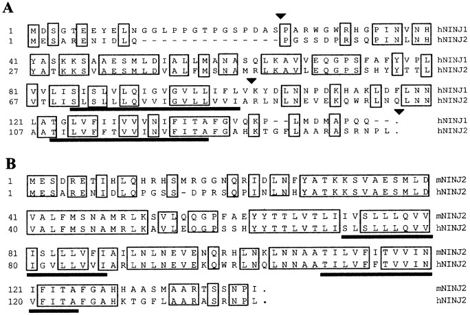
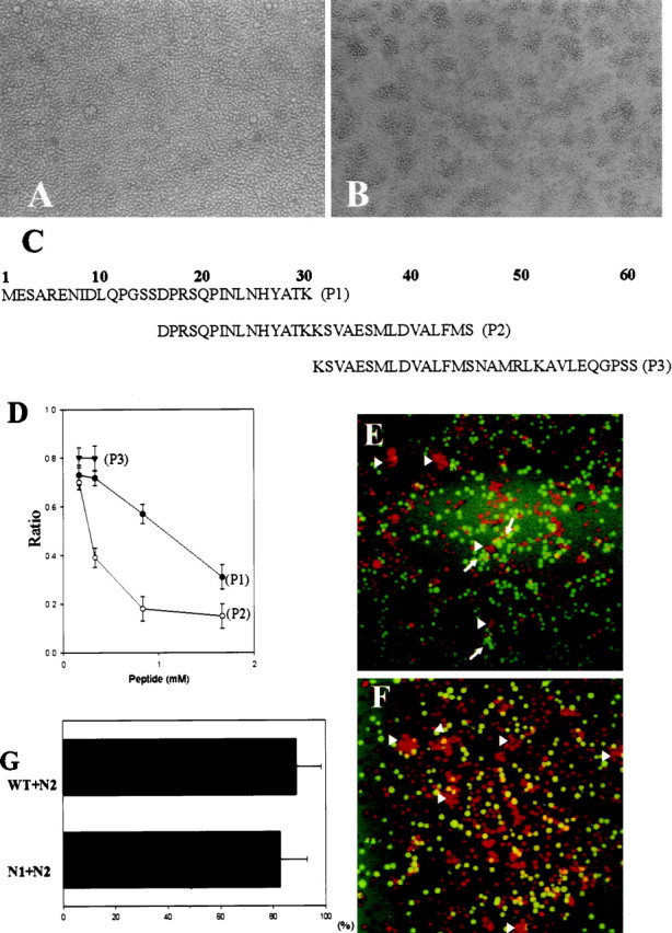
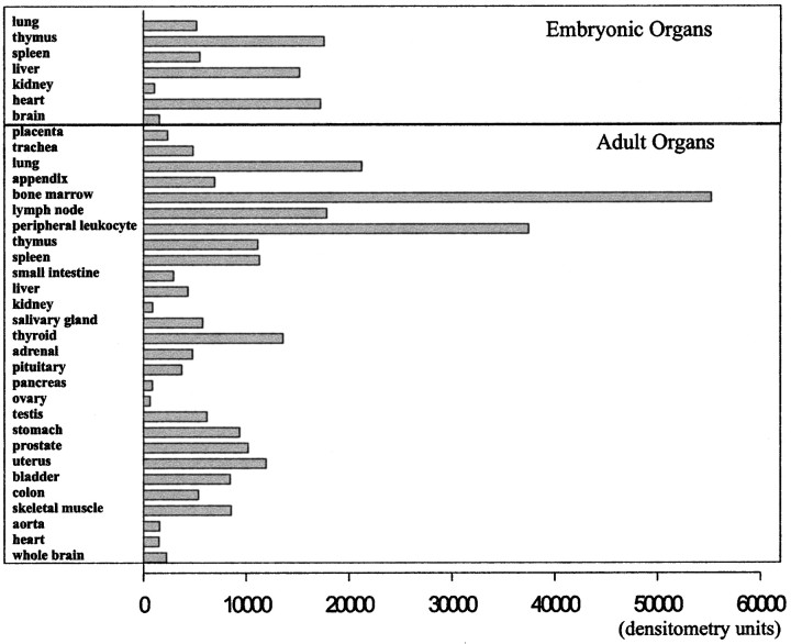
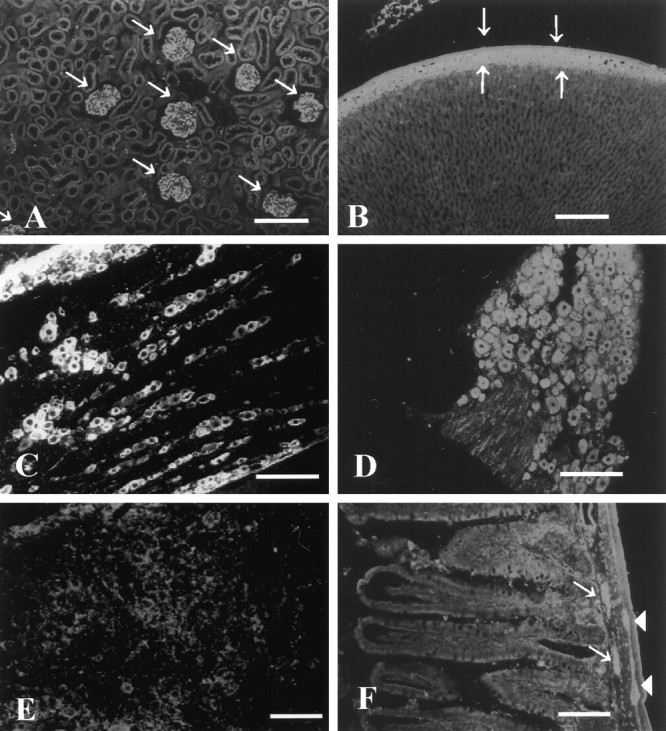
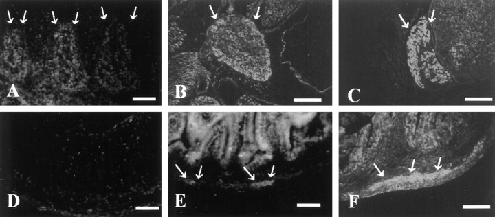
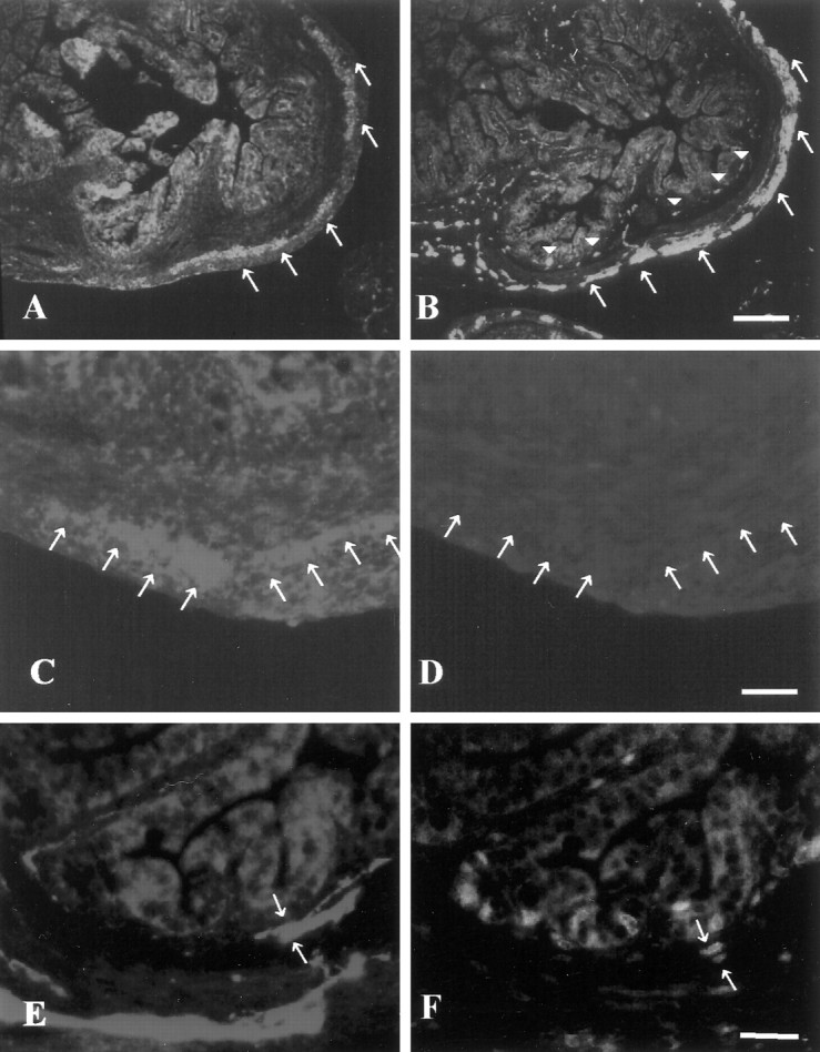
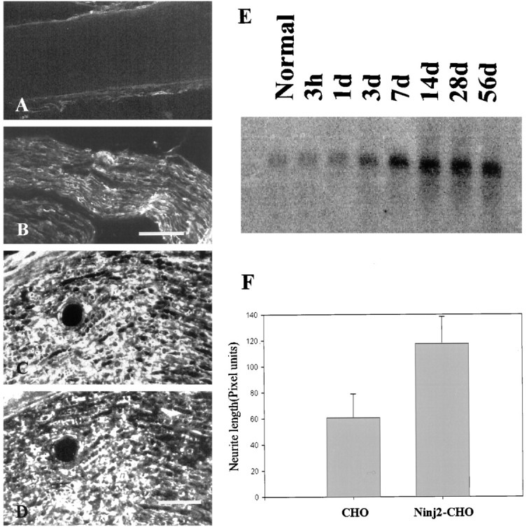
A large number of cell adhesion molecules mediate cell-to-cell and cell-to-extracellular matrix interaction during development, differentiation and regeneration of the peripheral nervous system. Here, we report the identification of a novel cell surface adhesion molecule, ninjurin2 (for nerve injury induced protein 2). Ninjurin2 is a homolog of a homophilic cellular adhesion molecule, ninjurin1, that was previously isolated as a gene induced in Schwann cells after nerve injury. Ninjurin1 and 2 share conserved hydrophobic regions for their transmembrane domains; however, they do not contain comparable adhesion motifs nor do they interact with each other. In the peripheral nervous system, ninjurin2 is expressed constitutively in mature sensory and enteric neurons but not in glial cells or in autonomic ganglia. Ninjurin2 is upregulated in Schwann cells surrounding the distal segment of injured nerve with a time course similar to that of ninjurin1, neural CAM, and L1. Ninjurin2 promotes neurite outgrowth from primary cultured dorsal root ganglion neurons, presumably via homophilic cellular interactions. Ninjurin2 is also highly expressed in hematopoietic and lymphatic tissues. Finally, the ninjurin2 gene is located on human chromosome 12p13 in which several disorders of unknown etiology have been mapped, including inflammatory bowel disease and acrocallosal syndrome.


Figures

Fig. 1.
Fig. 1.
Sequence analysis of ninjurin2. A, Alignment of human ninjurin1 and ninjurin2. Identical residues areboxed, and putative transmembrane domains areunderlined. Arrowheads denote intron–exon junctions. Note that these sites are conserved between the ninjurin1 and 2 genes. B, Alignment of human and mouse ninjurin2 amino acid sequences. Identical residues areboxed, and putative transmembrane domains areunderlined.
Fig. 2.
Fig. 2.
Protein blot analysis of ninjurin2. Proteins from lysates prepared from native CHO cells (lane 1), CHO cells stably transfected with a pNINJ1 expression construct (lane 2), and CHO cells stably transfected with pNINJ2 construct (lane 3) were electrophoresed on 12% SDS-polyacrylamide gels, transferred to nitrocellulose, and incubated with affinity-purified anti-ninjurin2 antibodies. Ninjurin2 was visualized by using enhanced chemiluminescence.
Fig. 3.
Fig. 3.
Ninjurin2 is localized on the plasma membrane. Immunocytochemical localization of ninjurin2 on wild-type CHO cells (A) and CHO cells transfected with pNINJ2 (B–D). Anti-ninjurin2 antibodies were applied to cells either after fixation with 4% paraformaldehyde (A, B) or before fixation (C). In D, anti-ninjurin2 antisera were blocked by incubation with the peptide immunogen and applied to cells as in B (see Materials and Methods). Scale bar, 40 μm.
Fig. 4.
Fig. 4.
Ninjurin2 mediates homophilic cell adhesion. Native Jurkat cells (A) and Jurkat cells stably transfected with pNINJ2 (B) were resuspended at 1 × 10⁶ cells/ml and allowed to aggregate at 37°C for 1 hr. Note the presence of large aggregates inB. C, List of peptides used in competition experiments and also as antigens to raise anti-ninjurin2 antisera. D, Aggregation assays were performed using ninjurin2 stably expressing Jurkat cells in the presence of each of the indicated peptides. The number of cells in aggregates was determined after 1 hr. The ratio of cells in aggregates to total cells was calculated and plotted versus peptide concentration. Data represents the mean ± SD of three independent experiments. E,F, Aggregation assays were performed with a mixture of N2 cells (stained with red-orange fluorescent dye) and either N1 cells (E) or wild-type cells (F) (stained with greenfluorescent dye). Note that the green cells indicated byarrows and red-orange cells indicated byarrowheads individually form aggregates inE, and only red cells form aggregates inF as indicated by arrowheads.G, Quantitative analysis of aggregation assays using mixed cell populations. The aggregation assays were performed as inE and F, and red cells and green cells in each aggregate were counted. The graph represents the mean ± SD percentage of N2 cells in aggregates that consist predominantly of N2 cells.
Fig. 5.
Fig. 5.
Expression of ninjurin2 mRNA in human tissues. A human mRNA dot blot (Clontech) was probed with a32P-labeled fragment of the human ninjurin2 cDNA. The hybridization signal intensity was quantified by using a PhosphorImager.
Fig. 6.
Fig. 6.
Immunohistochemical analysis of ninjurin2 expression in adult rat. Affinity-purified antibodies were used to detect ninjurin2 expression by immunohistochemistry in kidney (A), adrenal (B), trigeminal ganglion (C), DRG (D), superior cervical ganglion (E), and enteric plexus (F). Arrows denote glomeruli inA and glomerular layer in the adrenal cortex inB. Arrows and arrowheadsdenote submucosal and myenteric plexus, respectively, inF. Scale bars, 100 μm.
Fig. 7.
Fig. 7.
Expression of ninjurin2 in sensory and enteric neurons during development. Immunohistochemistry was used to detect ninjurin2 in mouse DRG at E14 (A), E19 (B), and P3 (C) and in mouse enteric plexus at E17 (D), P1 (E), and P3 (F).Arrows denote the ganglia in each micrograph. Scale bars: A, D, E, 50 μm;B, C, F, 150 μm.
Fig. 8.
Fig. 8.
Ninjurin2 is expressed in postmitotic neurons in enteric ganglia. A, B, Ninjurin2 immunoreactivity in enteric plexus in P3 mouse gut (A) was compared with neuron-specific enolase immunoreactivity on an adjacent section (B). Note that myenteric neurons denoted by arrows express ninjurin2, but submucosal neurons denoted by arrowheadsin B, which differentiate later than myenteric neurons, lack ninjurin2 expression in A. Scale bar, 100 μm.C–F, BrdU and either ninjurin2 (C,D) or NSE (E, F) were visualized on the same section of mouse gut at P3. A mouse (P3) was injected with BrdU and killed 1 hr later for immunohistochemistry. Ninjurin2 (C) and NSE expression (E) were visualized by Cy3-conjugated secondary antibody, and proliferating cells were visualized by FITC-conjugated anti-BrdU immunohistochemistry (D,F). The ninjurin2-positive myenteric ganglia lack BrdU staining (arrows in C andD), whereas the NSE-positive cells in the submucosal ganglia indicated by arrows in E and Fare BrdU-positive. Note that C and Drepresent the same section as do E and F. Scale bars, 50 μm.
Fig. 9.
Fig. 9.
Ninjurin2 is upregulated in Schwann cells after nerve injury. A–D, Immunohistochemistry was used to detect ninjurin2 on a longitudinal section of the normal sciatic nerve (A) or sciatic nerve segment distal to the site of injury (7 d after injury; B, low magnification;D, high magnification). In C, the section adjacent to D was stained with antibodies to ninjurin1. The very similar expression patterns in C andD indicate that ninjurin2 is expressed in Schwann cells in injured nerve. Scale bars: B, 100 μm;D, 50 μm. E, Total RNA (10 μg) was isolated from sciatic nerves distal to the site of transection at the indicated times after axotomy. The samples were electrophoresed, blotted, and hybridized with a 32P-labeled ninjurin2 cDNA probe. F, Ninjurin2 promotes neurite outgrowth from primary cultured DRG neurons. DRG neurons from E16 rat embryos were seeded onto a confluent monolayer of control CHO cells or CHO cells expressing ninjurin2. Six hours later, the cells were fixed, and neurites were visualized by immunostaining with neurofilament H antibodies. Neurite length was quantified by measuring neurites from ∼50 neurons grown under each condition in three independent experiments. Data represent mean ± SD length.

Similar articles

Cited by

References

    1. Altschul SF, Gish W, Miller W, Myers EW, Lipman DJ. Basic local alignment search tool. J Mol Biol. 1990;215:403–410. - PubMed
    1. Araki T, Milbrandt J. Ninjurin, a novel adhesion molecule, is induced by nerve injury and promotes axonal growth. Neuron. 1996;17:353–361. - PubMed
    1. Araki T, Zimonjic DB, Popescu NC, Milbrandt J. Mechanism of homophilic binding mediated by ninjurin, a novel widely expressed adhesion molecule. J Biol Chem. 1997;272:21373–21380. - PubMed
    1. Barnes WM. PCR amplification of up to 35-kb DNA with high fidelity and high yield from lambda bacteriophage templates. Proc Natl Acad Sci USA. 1994;91:2216–2220. - PMC - PubMed
    1. Bixby JL, Lilien J, Reichardt LF. Identification of the major proteins that promote neural process outgrowth on Schwann cells in vitro. J Cell Biol. 1988;107:353–361. - PMC - PubMed

Publication types

MeSH terms