Mast cells migrate from blood to brain
- PMID: 10627616
- PMCID: PMC6774132
- DOI: 10.1523/JNEUROSCI.20-01-00401.2000
Mast cells migrate from blood to brain
Abstract
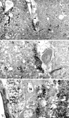
It is well established that mast cells (MCs) occur within the CNS of many species. Furthermore, their numbers can increase rapidly in adults in response to altered physiological conditions. In this study we found that early postpartum rats had significantly more mast cells in the thalamus than virgin controls. Evidence from semithin sections from these females suggested that mast cells were transiting across the medium-sized blood vessels. We hypothesized that the increases in mast cell number were caused by their migration into the neural parenchyma. To this end, we purified rat peritoneal mast cells, labeled them with the vital dyes PKH26 or CellTracker Green, and injected them into host animals. One hour after injection, dye-filled cells, containing either histamine or serotonin (mediators stored in mast cells), were located close to thalamic blood vessels. Injected cells represented approximately 2-20% of the total mast cell population in this brain region. Scanning confocal microscopy confirmed that the biogenic amine and the vital dye occurred in the same cell. To determine whether the donor mast cells were within the blood-brain barrier, we studied the localization of dye-marked donor cells and either Factor VIII, a component of endothelial basal laminae, or glial fibrillary acidic protein, the intermediate filament found in astrocytes. Serial section reconstructions of confocal images demonstrated that the mast cells were deep to the basal lamina, in nests of glial processes. This is the first demonstration that mast cells can rapidly penetrate brain blood vessels, and this may account for the rapid increases in mast cell populations after physiological manipulations.
Figures


References
-
- Abercrombie M. Estimation of nuclear population from microtomic sections. Anat Rec. 1946;94:239–247. - PubMed
-
- Ader R, Felten DL, Cohen N. Interactions between the brain and the immune system. Annu Rev Pharmacol Toxicol. 1990;30:561–602. - PubMed
-
- Airaksinen MS, Panula P. The histaminergic system in the guinea pig central nervous system: an immunocytochemical mapping study using an antiserum against histamine. J Comp Neurol. 1988;273:163–186. - PubMed
-
- Bacallao R, Kiai K, Jesaitis L. Guiding principles in specimen preservation for confocal microscopy. In: Pawley JB, editor. Handbook of biological confocal microscopy. Plenum; New York: 1995. pp. 311–326.
-
- Bacci S, Arbi-Riccardi R, Mayer B, Rumio C, Borghi-Cirri MB. Localization of nitric oxide synthetase immunoreactivity in mast cells of human nasal mucosa. Histochemistry. 1994;102:89–92. - PubMed
Publication types
MeSH terms
Substances
Grants and funding
LinkOut - more resources
Full Text Sources
Other Literature Sources
